Catalysis by Glomerella cingulata Cutinase Requires Conformational Cycling between the Active and Inactive States of Its Catalytic Triad
Nyon, M.P., Rice, D.W., Berrisford, J.M., Hounslow, A.M., Moir, A.J.G., Huang, H., Nathan, S., Mahadi, N.M., Bakar, F.D.A., Craven, C.J.(2009) J Mol Biol 385: 226-235
- PubMed: 18983850
- DOI: https://doi.org/10.1016/j.jmb.2008.10.050
- Primary Citation of Related Structures:
3DCN, 3DD5, 3DEA - PubMed Abstract:
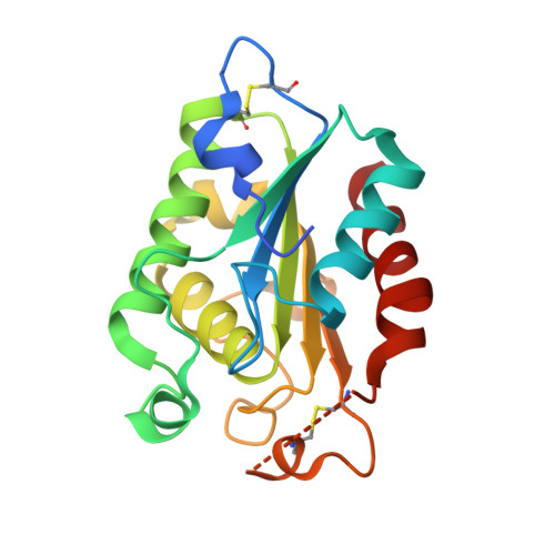
Cutinase belongs to a group of enzymes that catalyze the hydrolysis of esters and triglycerides. Structural studies on the enzyme from Fusarium solani have revealed the presence of a classic catalytic triad that has been implicated in the enzyme's mechanism. We have solved the crystal structure of Glomerella cingulata cutinase in the absence and in the presence of the inhibitors E600 (diethyl p-nitrophenyl phosphate) and PETFP (3-phenethylthio-1,1,1-trifluoropropan-2-one) to resolutions between 2.6 and 1.9 A. Analysis of these structures reveals that the catalytic triad (Ser136, Asp191, and His204) adopts an unusual configuration with the putative essential histidine His204 swung out of the active site into a position where it is unable to participate in catalysis, with the imidazole ring 11 A away from its expected position. Solution-state NMR experiments are consistent with the disrupted configuration of the triad observed crystallographically. H204N, a site-directed mutant, was shown to be catalytically inactive, confirming the importance of this residue in the enzyme mechanism. These findings suggest that, during its catalytic cycle, cutinase undergoes a significant conformational rearrangement converting the loop bearing the histidine from an inactive conformation, in which the histidine of the triad is solvent exposed, to an active conformation, in which the triad assumes a classic configuration.
Organizational Affiliation:
School of Biosciences and Biotechnology, Faculty of Science and Technology, Universiti Kebangsaan Malaysia, Selangor, Malaysia.