Structural analysis of substrate and effector binding in Mycobacterium tuberculosis D-3-phosphoglycerate dehydrogenase
Dey, S., Burton, R.L., Grant, G.A., Sacchettini, J.C.(2008) Biochemistry 47: 8271-8282
- PubMed: 18627175
- DOI: https://doi.org/10.1021/bi800212b
- Primary Citation of Related Structures:
3DC2, 3DDN - PubMed Abstract:
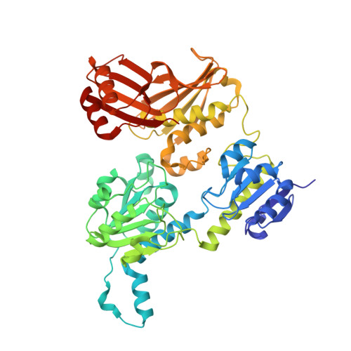
The crystal structure of Mycobacterium tuberculosis d-3-phosphoglycerate dehydrogenase has been solved with bound effector, l-serine, and substrate, hydroxypyruvic acid phosphate, at resolutions of 2.7 and 2.4 A, respectively. The subunits display the same extreme asymmetry as seen in the apo-structure and provide insight into the mode of serine binding and closure of the active site. Mutagenesis studies confirm the identity of the main residues involved in serine binding and suggest that the poly glycine stretch in the loop that contains the locus for the 160 degrees rotation that leads to subunit asymmetry may have a larger role in folding than in catalysis. The lack of electron density for the cofactor, NADH, in any of the crystals examined led us to study binding by stopped flow kinetic analysis. The kinetic data suggest that productive NADH binding, that would support catalytic turnover, is dependent on the presence of substrate. This observation, along with the binding of substrate in the active site, but in an unproductive conformation, suggests a possible mechanism where initial binding of substrate leads to enhanced interaction with cofactor accompanied by a rearrangement of catalytically critical residue side chains. Furthermore, comparison to the structure of a truncated form of human d-3-phosphoglycerate dehydrogenase with cofactor and a substrate analog, provides insight into the conformational changes that occur during catalysis.
Organizational Affiliation:
Department of Biochemistry & Biophysics, Texas A&M University, College Station, Texas 77843, USA.