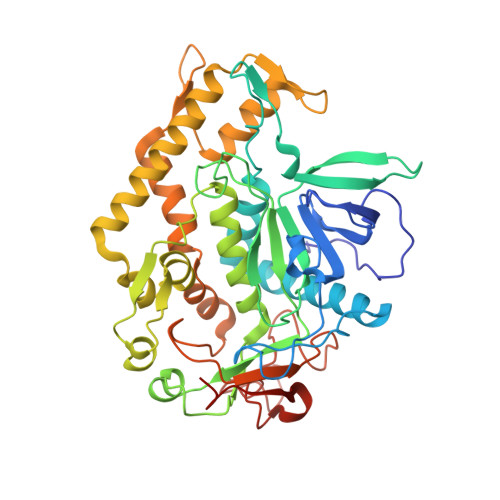
Substrate binding mode and its implication on drug design for botulinum neurotoxin A
Kumaran, D., Rawat, R., Ahmed, S.A., Swaminathan, S.(2008) PLoS Pathog 4: e1000165-e1000165
- PubMed: 18818739
- DOI: https://doi.org/10.1371/journal.ppat.1000165
- Primary Citation of Related Structures:
3DDA, 3DDB - PubMed Abstract:
The seven antigenically distinct serotypes of Clostridium botulinum neurotoxins, the causative agents of botulism, block the neurotransmitter release by specifically cleaving one of the three SNARE proteins and induce flaccid paralysis. The Centers for Disease Control and Prevention (CDC) has declared them as Category A biowarfare agents. The most potent among them, botulinum neurotoxin type A (BoNT/A), cleaves its substrate synaptosome-associated protein of 25 kDa (SNAP-25). An efficient drug for botulism can be developed only with the knowledge of interactions between the substrate and enzyme at the active site. Here, we report the crystal structures of the catalytic domain of BoNT/A with its uncleavable SNAP-25 peptide (197)QRATKM(202) and its variant (197)RRATKM(202) to 1.5 A and 1.6 A, respectively. This is the first time the structure of an uncleavable substrate bound to an active botulinum neurotoxin is reported and it has helped in unequivocally defining S1 to S5' sites. These substrate peptides make interactions with the enzyme predominantly by the residues from 160, 200, 250 and 370 loops. Most notably, the amino nitrogen and carbonyl oxygen of P1 residue (Gln197) chelate the zinc ion and replace the nucleophilic water. The P1'-Arg198, occupies the S1' site formed by Arg363, Thr220, Asp370, Thr215, Ile161, Phe163 and Phe194. The S2' subsite is formed by Arg363, Asn368 and Asp370, while S3' subsite is formed by Tyr251, Leu256, Val258, Tyr366, Phe369 and Asn388. P4'-Lys201 makes hydrogen bond with Gln162. P5'-Met202 binds in the hydrophobic pocket formed by the residues from the 250 and 200 loop. Knowledge of interactions between the enzyme and substrate peptide from these complex structures should form the basis for design of potent inhibitors for this neurotoxin.
Organizational Affiliation:
Biology Department, Brookhaven National Laboratory, Upton, New York, USA.