3DBO

Crystal structure of a member of the VapBC family of toxin-antitoxin systems, VapBC-5, from Mycobacterium tuberculosis


Experimental Data Snapshot

  • Method: X-RAY DIFFRACTION
  • Resolution: 1.76 Å
  • R-Value Free: 0.247 
  • R-Value Work: 0.213 
  • R-Value Observed: 0.214 

wwPDB Validation   3D Report Full Report


This is version 1.2 of the entry. See complete history


Literature

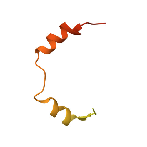
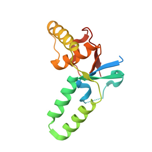
Structure and Proposed Activity of a Member of the VapBC Family of Toxin-Antitoxin Systems: VapBC-5 FROM MYCOBACTERIUM TUBERCULOSIS.

Miallau, L.Faller, M.Chiang, J.Arbing, M.Guo, F.Cascio, D.Eisenberg, D.

(2009) J Biol Chem 284: 276-283

  • DOI: https://doi.org/10.1074/jbc.M805061200
  • Primary Citation of Related Structures:  
    3DBO

  • PubMed Abstract: 

    In prokaryotes, cognate toxin-antitoxin pairs have long been known, but no three-dimensional structure has been available for any given complex from Mycobacterium tuberculosis. Here we report the crystal structure and activity of a member of the VapBC family of complexes from M. tuberculosis. The toxin VapC-5 is a compact, 150 residues, two domain alpha/beta protein. Bent around the toxin is the VapB-5 antitoxin, a 33-residue alpha-helix. Assays suggest that the toxin is an Mg-enabled endoribonuclease, inhibited by the antitoxin. The lack of DNase activity is consistent with earlier suggestions that the complex represses its own operon. Furthermore, analysis of the interactions in the binding of the antitoxin to the toxin suggest that exquisite control is required to protect the bacteria cell from toxic VapC-5.


  • Organizational Affiliation

    UCLA-DOE Institute of Genomics and Proteomics, the Department of Biological Chemistry, David Geffen School of Medicine, Molecular Cell and Developmental Biology, and the Department of Chemistry and Biochemistry, University of California, Los Angeles, Los Angeles, California 90095-1570.


Macromolecules
Find similar proteins by:  (by identity cutoff)  |  3D Structure
Entity ID: 1
MoleculeChains Sequence LengthOrganismDetailsImage
Uncharacterized protein93Mycobacterium tuberculosis H37RvMutation(s): 0 
Gene Names: Rv0626MT0654
UniProt
Find proteins for P9WF19 (Mycobacterium tuberculosis (strain ATCC 25618 / H37Rv))
Explore P9WF19 
Go to UniProtKB:  P9WF19
Entity Groups  
Sequence Clusters30% Identity50% Identity70% Identity90% Identity95% Identity100% Identity
UniProt GroupP9WF19
Sequence Annotations
Expand
  • Reference Sequence
Find similar proteins by:  (by identity cutoff)  |  3D Structure
Entity ID: 2
MoleculeChains Sequence LengthOrganismDetailsImage
Uncharacterized protein150Mycobacterium tuberculosis H37RvMutation(s): 0 
Gene Names: Rv0627
UniProt
Find proteins for P96917 (Mycobacterium tuberculosis (strain ATCC 25618 / H37Rv))
Explore P96917 
Go to UniProtKB:  P96917
Entity Groups  
Sequence Clusters30% Identity50% Identity70% Identity90% Identity95% Identity100% Identity
UniProt GroupP96917
Sequence Annotations
Expand
  • Reference Sequence
Experimental Data & Validation

Experimental Data

  • Method: X-RAY DIFFRACTION
  • Resolution: 1.76 Å
  • R-Value Free: 0.247 
  • R-Value Work: 0.213 
  • R-Value Observed: 0.214 
  • Space Group: P 65 2 2
Unit Cell:
Length ( Å )Angle ( ˚ )
a = 64.83α = 90
b = 64.83β = 90
c = 164.65γ = 120
Software Package:
Software NamePurpose
REFMACrefinement
HKL-2000data collection
DENZOdata reduction
SCALEPACKdata scaling
SHELXDphasing

Structure Validation

View Full Validation Report


Entry History 

Revision History  (Full details and data files)

  • Version 1.0: 2008-07-15
    Type: Initial release
  • Version 1.1: 2011-07-13
    Changes: Source and taxonomy, Version format compliance
  • Version 1.2: 2024-02-21
    Changes: Data collection, Database references, Derived calculations