3D9S

Human Aquaporin 5 (AQP5) - High Resolution X-ray Structure


Experimental Data Snapshot

  • Method: X-RAY DIFFRACTION
  • Resolution: 2.00 Å
  • R-Value Free: 0.193 
  • R-Value Work: 0.162 

wwPDB Validation   3D Report Full Report


Ligand Structure Quality Assessment 


This is version 1.2 of the entry. See complete history


Literature

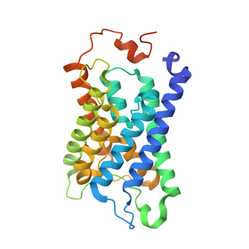
High-resolution x-ray structure of human aquaporin 5

Horsefield, R.Norden, K.Fellert, M.Backmark, A.Tornroth-Horsefield, S.Terwisscha van Scheltinga, A.C.Kvassman, J.Kjellbom, P.Johanson, U.Neutze, R.

(2008) Proc Natl Acad Sci U S A 105: 13327-13332

  • DOI: https://doi.org/10.1073/pnas.0801466105
  • Primary Citation of Related Structures:  
    3D9S

  • PubMed Abstract: 

    Human aquaporin 5 (HsAQP5) facilitates the transport of water across plasma membranes and has been identified within cells of the stomach, duodenum, pancreas, airways, lungs, salivary glands, sweat glands, eyes, lacrimal glands, and the inner ear. AQP5, like AQP2, is subject to posttranslational regulation by phosphorylation, at which point it is trafficked between intracellular storage compartments and the plasma membrane. Details concerning the molecular mechanism of membrane trafficking are unknown. Here we report the x-ray structure of HsAQP5 to 2.0-A resolution and highlight structural similarities and differences relative to other eukaryotic aquaporins. A lipid occludes the putative central pore, preventing the passage of gas or ions through the center of the tetramer. Multiple consensus phosphorylation sites are observed in the structure and their potential regulatory role is discussed. We postulate that a change in the conformation of the C terminus may arise from the phosphorylation of AQP5 and thereby signal trafficking.


  • Organizational Affiliation

    Department of Chemistry, Biochemistry and Biophysics, University of Gothenburg, Box 462, SE-405 30 Göteborg, Sweden.


Macromolecules
Find similar proteins by:  (by identity cutoff)  |  3D Structure
Entity ID: 1
MoleculeChains Sequence LengthOrganismDetailsImage
Aquaporin-5
A, B, C, D
266Homo sapiensMutation(s): 0 
Gene Names: AQP5
Membrane Entity: Yes 
UniProt & NIH Common Fund Data Resources
Find proteins for P55064 (Homo sapiens)
Explore P55064 
Go to UniProtKB:  P55064
PHAROS:  P55064
GTEx:  ENSG00000161798 
Entity Groups  
Sequence Clusters30% Identity50% Identity70% Identity90% Identity95% Identity100% Identity
UniProt GroupP55064
Sequence Annotations
Expand
  • Reference Sequence
Small Molecules
Ligands 1 Unique
IDChains Name / Formula / InChI Key2D Diagram3D Interactions
PS6
Query on PS6

Download Ideal Coordinates CCD File 
E [auth D]O-[(S)-{[(2S)-2-(hexanoyloxy)-3-(tetradecanoyloxy)propyl]oxy}(hydroxy)phosphoryl]-D-serine
C26 H50 N O10 P
HXSPCZGHXODQMW-XZOQPEGZSA-N
Experimental Data & Validation

Experimental Data

  • Method: X-RAY DIFFRACTION
  • Resolution: 2.00 Å
  • R-Value Free: 0.193 
  • R-Value Work: 0.162 
  • Space Group: P 21 21 21
Unit Cell:
Length ( Å )Angle ( ˚ )
a = 90.477α = 90
b = 90.644β = 90
c = 184.395γ = 90
Software Package:
Software NamePurpose
SHELXmodel building
SHELXL-97refinement
MxCuBEdata collection
MOSFLMdata reduction
SCALAdata scaling
AMoREphasing

Structure Validation

View Full Validation Report


Ligand Structure Quality Assessment 


Entry History 

Revision History  (Full details and data files)

  • Version 1.0: 2008-08-26
    Type: Initial release
  • Version 1.1: 2011-07-13
    Changes: Version format compliance
  • Version 1.2: 2023-11-01
    Changes: Data collection, Database references, Derived calculations, Refinement description