3D98

Crystal structure of GlmU from Mycobacterium tuberculosis, ligand-free form


Experimental Data Snapshot

  • Method: X-RAY DIFFRACTION
  • Resolution: 2.50 Å
  • R-Value Free: 0.244 
  • R-Value Work: 0.197 
  • R-Value Observed: 0.199 

wwPDB Validation   3D Report Full Report


This is version 1.3 of the entry. See complete history


Literature

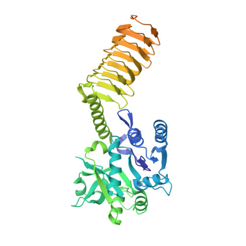
Structure and function of GlmU from Mycobacterium tuberculosis.

Zhang, Z.Bulloch, E.M.Bunker, R.D.Baker, E.N.Squire, C.J.

(2009) Acta Crystallogr D Biol Crystallogr 65: 275-283

  • DOI: https://doi.org/10.1107/S0907444909001036
  • Primary Citation of Related Structures:  
    2QKX, 3D8V, 3D98

  • PubMed Abstract: 

    Antibiotic resistance is a major issue in the treatment of infectious diseases such as tuberculosis. Existing antibiotics target only a few cellular pathways and there is an urgent need for antibiotics that have novel molecular mechanisms. The glmU gene is essential in Mycobacterium tuberculosis, being required for optimal bacterial growth, and has been selected as a possible drug target for structural and functional investigation. GlmU is a bifunctional acetyltransferase/uridyltransferase that catalyses the formation of UDP-GlcNAc from GlcN-1-P. UDP-GlcNAc is a substrate for two important biosynthetic pathways: lipopolysaccharide and peptidoglycan synthesis. The crystal structure of M. tuberculosis GlmU has been determined in an unliganded form and in complex with GlcNAc-1-P or UDP-GlcNAc. The structures reveal the residues that are responsible for substrate binding. Enzyme activities were characterized by (1)H NMR and suggest that the presence of acetyl-coenzyme A has an inhibitory effect on uridyltransferase activity.


  • Organizational Affiliation

    School of Biological Sciences and Maurice Wilkins Centre for Molecular Biodiscovery, University of Auckland, New Zealand.


Macromolecules
Find similar proteins by:  (by identity cutoff)  |  3D Structure
Entity ID: 1
MoleculeChains Sequence LengthOrganismDetailsImage
Bifunctional protein glmU495Mycobacterium tuberculosisMutation(s): 0 
Gene Names: glmURv1018cMT1046
EC: 2.7.7.23 (PDB Primary Data), 2.3.1.157 (PDB Primary Data)
UniProt
Find proteins for P9WMN3 (Mycobacterium tuberculosis (strain ATCC 25618 / H37Rv))
Explore P9WMN3 
Go to UniProtKB:  P9WMN3
Entity Groups  
Sequence Clusters30% Identity50% Identity70% Identity90% Identity95% Identity100% Identity
UniProt GroupP9WMN3
Sequence Annotations
Expand
  • Reference Sequence
Experimental Data & Validation

Experimental Data

  • Method: X-RAY DIFFRACTION
  • Resolution: 2.50 Å
  • R-Value Free: 0.244 
  • R-Value Work: 0.197 
  • R-Value Observed: 0.199 
  • Space Group: H 3 2
Unit Cell:
Length ( Å )Angle ( ˚ )
a = 94.27α = 90
b = 94.27β = 90
c = 284.226γ = 120
Software Package:
Software NamePurpose
PHENIXrefinement
Blu-Icedata collection
MOSFLMdata reduction
SCALAdata scaling
PHASERphasing

Structure Validation

View Full Validation Report


Entry History 

Deposition Data

Revision History  (Full details and data files)

  • Version 1.0: 2009-03-10
    Type: Initial release
  • Version 1.1: 2011-07-13
    Changes: Version format compliance
  • Version 1.2: 2017-10-25
    Changes: Refinement description
  • Version 1.3: 2023-08-30
    Changes: Data collection, Database references, Refinement description