3D7U

Structural basis for the recognition of c-Src by its inactivator Csk


Experimental Data Snapshot

  • Method: X-RAY DIFFRACTION
  • Resolution: 4.11 Å
  • R-Value Free: 0.302 
  • R-Value Work: 0.294 
  • R-Value Observed: 0.294 

wwPDB Validation   3D Report Full Report


This is version 1.3 of the entry. See complete history


Literature

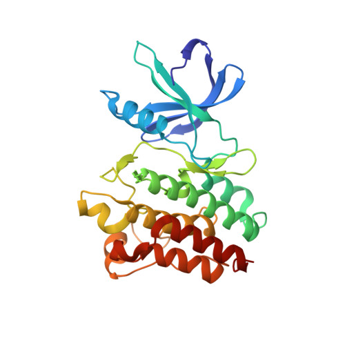
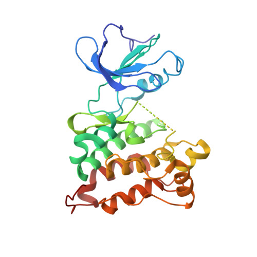
Structural basis for the recognition of c-Src by its inactivator Csk.

Levinson, N.M.Seeliger, M.A.Cole, P.A.Kuriyan, J.

(2008) Cell 134: 124-134

  • DOI: https://doi.org/10.1016/j.cell.2008.05.051
  • Primary Citation of Related Structures:  
    3D7T, 3D7U

  • PubMed Abstract: 

    The catalytic activity of the Src family of tyrosine kinases is suppressed by phosphorylation on a tyrosine residue located near the C terminus (Tyr 527 in c-Src), which is catalyzed by C-terminal Src Kinase (Csk). Given the promiscuity of most tyrosine kinases, it is remarkable that the C-terminal tails of the Src family kinases are the only known targets of Csk. We have determined the crystal structure of a complex between the kinase domains of Csk and c-Src at 2.9 A resolution, revealing that interactions between these kinases position the C-terminal tail of c-Src at the edge of the active site of Csk. Csk cannot phosphorylate substrates that lack this docking mechanism because the conventional substrate binding site used by most tyrosine kinases to recognize substrates is destabilized in Csk by a deletion in the activation loop.


  • Organizational Affiliation

    Department of Molecular and Cell Biology, Department of Chemistry, Howard Hughes Medical Institute, California Institute for Quantitative Biosciences (QB3), University of California, Berkeley, Berkeley, CA 94720, USA.


Macromolecules
Find similar proteins by:  (by identity cutoff)  |  3D Structure
Entity ID: 1
MoleculeChains Sequence LengthOrganismDetailsImage
Tyrosine-protein kinase CSK
A, C
263Homo sapiensMutation(s): 0 
Gene Names: CSK
EC: 2.7.10.2
UniProt & NIH Common Fund Data Resources
Find proteins for P41240 (Homo sapiens)
Explore P41240 
Go to UniProtKB:  P41240
PHAROS:  P41240
GTEx:  ENSG00000103653 
Entity Groups  
Sequence Clusters30% Identity50% Identity70% Identity90% Identity95% Identity100% Identity
UniProt GroupP41240
Sequence Annotations
Expand
  • Reference Sequence
Find similar proteins by:  (by identity cutoff)  |  3D Structure
Entity ID: 2
MoleculeChains Sequence LengthOrganismDetailsImage
Proto-oncogene tyrosine-protein kinase Src
B, D
277Gallus gallusMutation(s): 0 
Gene Names: SRC
EC: 2.7.10.2
UniProt
Find proteins for P00523 (Gallus gallus)
Explore P00523 
Go to UniProtKB:  P00523
Entity Groups  
Sequence Clusters30% Identity50% Identity70% Identity90% Identity95% Identity100% Identity
UniProt GroupP00523
Sequence Annotations
Expand
  • Reference Sequence
Experimental Data & Validation

Experimental Data

  • Method: X-RAY DIFFRACTION
  • Resolution: 4.11 Å
  • R-Value Free: 0.302 
  • R-Value Work: 0.294 
  • R-Value Observed: 0.294 
  • Space Group: P 31
Unit Cell:
Length ( Å )Angle ( ˚ )
a = 135.839α = 90
b = 135.839β = 90
c = 129.509γ = 120
Software Package:
Software NamePurpose
PHENIXrefinement
HKL-2000data reduction
HKL-2000data scaling

Structure Validation

View Full Validation Report


Entry History 

Deposition Data

Revision History  (Full details and data files)

  • Version 1.0: 2008-08-05
    Type: Initial release
  • Version 1.1: 2011-07-13
    Changes: Version format compliance
  • Version 1.2: 2013-09-18
    Changes: Derived calculations
  • Version 1.3: 2024-02-21
    Changes: Data collection, Database references