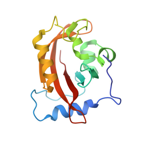
3D72

1.65 Angstrom crystal structure of the Cys71Val variant in the fungal photoreceptor VVD


Experimental Data Snapshot

  • Method: X-RAY DIFFRACTION
  • Resolution: 1.65 Å
  • R-Value Free: 0.260 
  • R-Value Work: 0.250 
  • R-Value Observed: 0.260 

wwPDB Validation   3D Report Full Report


Ligand Structure Quality Assessment 


This is version 1.3 of the entry. See complete history


Literature

Light activation of the LOV protein vivid generates a rapidly exchanging dimer.

Zoltowski, B.D.Crane, B.R.

(2008) Biochemistry 47: 7012-7019

  • DOI: https://doi.org/10.1021/bi8007017
  • Primary Citation of Related Structures:  
    3D72

  • PubMed Abstract: 

    The fungal photoreceptor Vivid (VVD) plays an important role in the adaptation of blue-light responses in Neurospora crassa. VVD, an FAD-binding LOV (light, oxygen, voltage) protein, couples light-induced cysteinyl adduct formation at the flavin ring to conformational changes in the N-terminal cap (Ncap) of the VVD PAS domain. Size-exclusion chromatography (SEC), equilibrium ultracentrifugation, and static and dynamic light scattering show that these conformational changes generate a rapidly exchanging VVD dimer, with an expanded hydrodynamic radius. A three-residue N-terminal beta-turn that assumes two different conformations in a crystal structure of a VVD C71V variant is essential for light-state dimerization. Residue substitutions at a critical hinge between the Ncap and PAS core can inhibit or enhance dimerization, whereas a Tyr to Trp substitution at the Ncap-PAS interface stabilizes the light-state dimer. Cross-linking through engineered disulfides indicates that the light-state dimer differs considerably from the dark-state dimer found in VVD crystal structures. These results verify the role of Ncap conformational changes in gating the photic response of N. crassa and indicate that LOV-LOV homo- or heterodimerization may be a mechanism for regulating light-activated gene expression.


  • Organizational Affiliation

    Department of Chemistry and Chemical Biology, Cornell University, Ithaca, New York 14853, USA.


Macromolecules
Find similar proteins by:  (by identity cutoff)  |  3D Structure
Entity ID: 1
MoleculeChains Sequence LengthOrganismDetailsImage
Vivid PAS protein VVD
A, B
149Neurospora crassaMutation(s): 1 
Gene Names: vvdG17A4.050
UniProt
Find proteins for Q1K5Y8 (Neurospora crassa (strain ATCC 24698 / 74-OR23-1A / CBS 708.71 / DSM 1257 / FGSC 987))
Explore Q1K5Y8 
Go to UniProtKB:  Q1K5Y8
Entity Groups  
Sequence Clusters30% Identity50% Identity70% Identity90% Identity95% Identity100% Identity
UniProt GroupQ1K5Y8
Sequence Annotations
Expand
  • Reference Sequence
Small Molecules
Ligands 1 Unique
IDChains Name / Formula / InChI Key2D Diagram3D Interactions
FAD
Query on FAD

Download Ideal Coordinates CCD File 
C [auth A],
D [auth B]
FLAVIN-ADENINE DINUCLEOTIDE
C27 H33 N9 O15 P2
VWWQXMAJTJZDQX-UYBVJOGSSA-N
Experimental Data & Validation

Experimental Data

  • Method: X-RAY DIFFRACTION
  • Resolution: 1.65 Å
  • R-Value Free: 0.260 
  • R-Value Work: 0.250 
  • R-Value Observed: 0.260 
  • Space Group: P 1 21 1
Unit Cell:
Length ( Å )Angle ( ˚ )
a = 36.24α = 90
b = 80.7β = 95.16
c = 57.58γ = 90
Software Package:
Software NamePurpose
HKL-2000data collection
AMoREphasing
CNSrefinement
HKL-2000data reduction
HKL-2000data scaling

Structure Validation

View Full Validation Report


Ligand Structure Quality Assessment 


Entry History 

Deposition Data

Revision History  (Full details and data files)

  • Version 1.0: 2008-06-17
    Type: Initial release
  • Version 1.1: 2011-07-13
    Changes: Version format compliance
  • Version 1.2: 2018-06-27
    Changes: Data collection, Database references
  • Version 1.3: 2023-08-30
    Changes: Data collection, Database references, Derived calculations, Refinement description