3D5K

Crystal structure of the OprM channel in a non-symmetrical space group


Experimental Data Snapshot

  • Method: X-RAY DIFFRACTION
  • Resolution: 2.40 Å
  • R-Value Free: 0.293 
  • R-Value Work: 0.236 
  • R-Value Observed: 0.240 

wwPDB Validation   3D Report Full Report


This is version 1.3 of the entry. See complete history


Literature

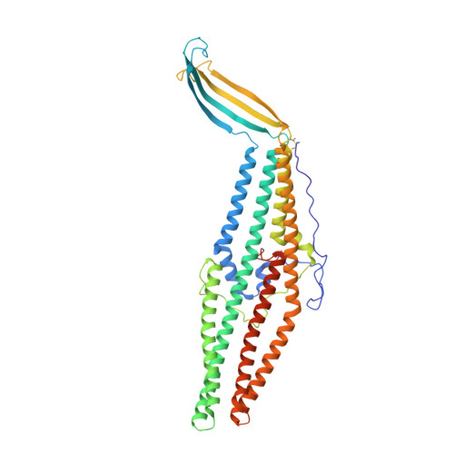
Structural and dynamical insights into the opening mechanism of P. aeruginosa OprM channel.

Phan, G.Benabdelhak, H.Lascombe, M.B.Benas, P.Rety, S.Picard, M.Ducruix, A.Etchebest, C.Broutin, I.

(2010) Structure 18: 507-517

  • DOI: https://doi.org/10.1016/j.str.2010.01.018
  • Primary Citation of Related Structures:  
    3D5K

  • PubMed Abstract: 

    Originally described in bacteria, drug transporters are now recognized as major determinants in antibiotics resistance. For Gram-negative bacteria, the reversible assembly consisting of an inner membrane protein responsible for the active transport, a periplasmic protein, and an exit outer membrane channel achieves transport. The opening of the outer membrane protein OprM from Pseudomonas aeruginosa was modeled through normal mode analysis starting from a new X-ray structure solved at 2.4 A resolution in P2(1)2(1)2(1) space group. The three monomers are not linked by internal crystallographic symmetries highlighting the possible functional differences. This structure is closed at both ends, but modeling allowed for an opening that is not reduced to the classically proposed "iris-like mechanism."


  • Organizational Affiliation

    Laboratoire de Cristallographie et RMN Biologiques, Université Paris Descartes, UMR 8015 CNRS, Faculté des Sciences Pharmaceutiques et Biologiques, 4 Avenue de l'Observatoire, 75270 Paris Cedex 06, France.


Macromolecules
Find similar proteins by:  (by identity cutoff)  |  3D Structure
Entity ID: 1
MoleculeChains Sequence LengthOrganismDetailsImage
Outer membrane protein oprM
A, B, C
474Pseudomonas aeruginosaMutation(s): 0 
Gene Names: oprMoprKPA0427
Membrane Entity: Yes 
UniProt
Find proteins for Q51487 (Pseudomonas aeruginosa (strain ATCC 15692 / DSM 22644 / CIP 104116 / JCM 14847 / LMG 12228 / 1C / PRS 101 / PAO1))
Explore Q51487 
Go to UniProtKB:  Q51487
Entity Groups  
Sequence Clusters30% Identity50% Identity70% Identity90% Identity95% Identity100% Identity
UniProt GroupQ51487
Sequence Annotations
Expand
  • Reference Sequence
Small Molecules
Ligands 2 Unique
IDChains Name / Formula / InChI Key2D Diagram3D Interactions
CL
Query on CL

Download Ideal Coordinates CCD File 
F [auth B]CHLORIDE ION
Cl
VEXZGXHMUGYJMC-UHFFFAOYSA-M
NA
Query on NA

Download Ideal Coordinates CCD File 
D [auth A]
E [auth A]
G [auth B]
H [auth B]
I [auth B]
D [auth A],
E [auth A],
G [auth B],
H [auth B],
I [auth B],
J [auth B],
K [auth B],
L [auth C],
M [auth C]
SODIUM ION
Na
FKNQFGJONOIPTF-UHFFFAOYSA-N
Modified Residues  1 Unique
IDChains TypeFormula2D DiagramParent
SCY
Query on SCY
A, B, C
L-PEPTIDE LINKINGC5 H9 N O3 SCYS
Experimental Data & Validation

Experimental Data

  • Method: X-RAY DIFFRACTION
  • Resolution: 2.40 Å
  • R-Value Free: 0.293 
  • R-Value Work: 0.236 
  • R-Value Observed: 0.240 
  • Space Group: P 21 21 21
Unit Cell:
Length ( Å )Angle ( ˚ )
a = 73.9α = 90
b = 70.2β = 90
c = 344.5γ = 90
Software Package:
Software NamePurpose
REFMACrefinement
ADSCdata collection
XDSdata reduction
XDSdata scaling
PHASERphasing

Structure Validation

View Full Validation Report


Entry History 

Deposition Data

Revision History  (Full details and data files)

  • Version 1.0: 2009-06-02
    Type: Initial release
  • Version 1.1: 2011-07-13
    Changes: Version format compliance
  • Version 1.2: 2012-04-18
    Changes: Database references
  • Version 1.3: 2023-11-01
    Changes: Data collection, Database references, Derived calculations, Refinement description