3D54

Structure of PurLQS from Thermotoga maritima


Experimental Data Snapshot

  • Method: X-RAY DIFFRACTION
  • Resolution: 3.50 Å
  • R-Value Free: 0.282 
  • R-Value Work: 0.252 
  • R-Value Observed: 0.252 

wwPDB Validation   3D Report Full Report


Ligand Structure Quality Assessment 


This is version 1.2 of the entry. See complete history


Literature

Formylglycinamide ribonucleotide amidotransferase from Thermotoga maritima: structural insights into complex formation.

Morar, M.Hoskins, A.A.Stubbe, J.Ealick, S.E.

(2008) Biochemistry 47: 7816-7830

  • DOI: https://doi.org/10.1021/bi800329p
  • Primary Citation of Related Structures:  
    3D54

  • PubMed Abstract: 

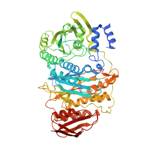
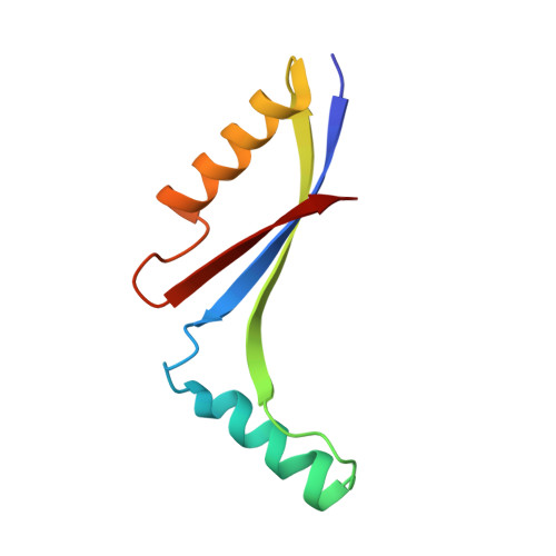
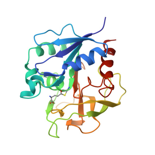
    In the fourth step of the purine biosynthetic pathway, formyl glycinamide ribonucleotide (FGAR) amidotransferase, also known as PurL, catalyzes the conversion of FGAR, ATP, and glutamine to formyl glycinamidine ribonucleotide (FGAM), ADP, P i, and glutamate. Two forms of PurL have been characterized, large and small. Large PurL, present in most Gram-negative bacteria and eukaryotes, consists of a single polypeptide chain and contains three major domains: the N-terminal domain, the FGAM synthetase domain, and the glutaminase domain, with a putative ammonia channel located between the active sites of the latter two. Small PurL, present in Gram-positive bacteria and archaea, is structurally homologous to the FGAM synthetase domain of large PurL, and forms a complex with two additional gene products, PurQ and PurS. The structure of the PurS dimer is homologous with the N-terminal domain of large PurL, while PurQ, whose structure has not been reported, contains the glutaminase activity. In Bacillus subtilis, the formation of the PurLQS complex is dependent on glutamine and ADP and has been demonstrated by size-exclusion chromatography. In this work, a structure of the PurLQS complex from Thermotoga maritima is described revealing a 2:1:1 stoichiometry of PurS:Q:L, respectively. The conformational changes observed in TmPurL upon complex formation elucidate the mechanism of metabolite-mediated recruitment of PurQ and PurS. The flexibility of the PurS dimer is proposed to play a role in the activation of the complex and the formation of the ammonia channel. A potential path for the ammonia channel is identified.


  • Organizational Affiliation

    Department of Chemistry and Chemical Biology, Cornell University, Ithaca, New York 14853, USA.


Macromolecules
Find similar proteins by:  (by identity cutoff)  |  3D Structure
Entity ID: 1
MoleculeChains Sequence LengthOrganismDetailsImage
Phosphoribosylformylglycinamidine synthase II
A, E, I
629Thermotoga maritimaMutation(s): 0 
Gene Names: purL
EC: 6.3.5.3
UniProt
Find proteins for Q9X0X3 (Thermotoga maritima (strain ATCC 43589 / DSM 3109 / JCM 10099 / NBRC 100826 / MSB8))
Explore Q9X0X3 
Go to UniProtKB:  Q9X0X3
Entity Groups  
Sequence Clusters30% Identity50% Identity70% Identity90% Identity95% Identity100% Identity
UniProt GroupQ9X0X3
Sequence Annotations
Expand
  • Reference Sequence
Find similar proteins by:  (by identity cutoff)  |  3D Structure
Entity ID: 2
MoleculeChains Sequence LengthOrganismDetailsImage
Formylglycinamide ribonucleotide amidotransferase
B, C, F, G, J
B, C, F, G, J, K
82Thermotoga maritimaMutation(s): 0 
EC: 6.3.5.3
UniProt
Find proteins for Q9X0X1 (Thermotoga maritima (strain ATCC 43589 / DSM 3109 / JCM 10099 / NBRC 100826 / MSB8))
Explore Q9X0X1 
Go to UniProtKB:  Q9X0X1
Entity Groups  
Sequence Clusters30% Identity50% Identity70% Identity90% Identity95% Identity100% Identity
UniProt GroupQ9X0X1
Sequence Annotations
Expand
  • Reference Sequence
Find similar proteins by:  (by identity cutoff)  |  3D Structure
Entity ID: 3
MoleculeChains Sequence LengthOrganismDetailsImage
Phosphoribosylformylglycinamidine synthase 1
D, H, L
213Thermotoga maritimaMutation(s): 0 
Gene Names: purQ
EC: 6.3.5.3
UniProt
Find proteins for Q9X0X2 (Thermotoga maritima (strain ATCC 43589 / DSM 3109 / JCM 10099 / NBRC 100826 / MSB8))
Explore Q9X0X2 
Go to UniProtKB:  Q9X0X2
Entity Groups  
Sequence Clusters30% Identity50% Identity70% Identity90% Identity95% Identity100% Identity
UniProt GroupQ9X0X2
Sequence Annotations
Expand
  • Reference Sequence
Experimental Data & Validation

Experimental Data

  • Method: X-RAY DIFFRACTION
  • Resolution: 3.50 Å
  • R-Value Free: 0.282 
  • R-Value Work: 0.252 
  • R-Value Observed: 0.252 
  • Space Group: C 1 2 1
Unit Cell:
Length ( Å )Angle ( ˚ )
a = 256.851α = 90
b = 187.337β = 99.12
c = 159.177γ = 90
Software Package:
Software NamePurpose
CNSrefinement
HKL-2000data collection
HKL-2000data reduction
HKL-2000data scaling
PHASERphasing

Structure Validation

View Full Validation Report


Ligand Structure Quality Assessment 


Entry History 

Deposition Data

Revision History  (Full details and data files)

  • Version 1.0: 2008-07-22
    Type: Initial release
  • Version 1.1: 2011-07-13
    Changes: Version format compliance
  • Version 1.2: 2017-02-01
    Changes: Structure summary