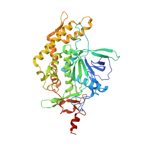
3D3X

Crystal structure of botulinum neurotoxin serotype E catalytic domain in complex with SNAP-25 substrate peptide


Experimental Data Snapshot

  • Method: X-RAY DIFFRACTION
  • Resolution: 2.25 Å
  • R-Value Free: 0.288 
  • R-Value Work: 0.235 
  • R-Value Observed: 0.235 

wwPDB Validation   3D Report Full Report


This is version 1.3 of the entry. See complete history


Literature

SNAP-25 substrate peptide (residues 180-183) binds to but bypasses cleavage by catalytically active Clostridium botulinum neurotoxin E.

Agarwal, R.Swaminathan, S.

(2008) J Biol Chem 283: 25944-25951

  • DOI: https://doi.org/10.1074/jbc.M803756200
  • Primary Citation of Related Structures:  
    3D3X

  • PubMed Abstract: 

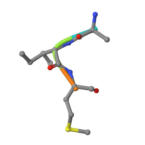
    Clostridium botulinum neurotoxins are the most potent toxins to humans. The recognition and cleavage of SNAREs are prime evente in exhibiting their toxicity. We report here the crystal structure of the catalytically active full-length botulinum serotype E catalytic domain (BoNT E) in complex with SNAP-25 (a SNARE protein) substrate peptide Arg(180)-Ile(181)-Met(182)-Glu(183) (P1-P3'). It is remarkable that the peptide spanning the scissile bond binds to but bypasses cleavage by the enzyme and inhibits the catalysis fairly with K(i) approximately 69 microm. The inhibitory peptide occupies the active site of BoNT E and shows well defined electron density. The catalytic zinc and the conserved key residue Tyr(350) of the enzyme facilitate the docking of Arg(180) (P1) by interacting with its carbonyl oxygen that displaces the nucleophilic water. The general base Glu(212) side chain interacts with the main chain amino group of P1 and P1'. Conserved Arg(347) of BoNT E stabilizes the proper docking of the Ile(181) (P1') main chain, whereas the hydrophobic pockets stabilize the side chains of Ile(181) (P1') and Met(182) (P2'), and the 250 loop stabilizes Glu(183) (P3'). Structural and functional analysis revealed an important role for the P1' residue and S1' pocket in driving substrate recognition and docking at the active site. This study is the first of its kind and rationalizes the substrate cleavage strategy of BoNT E. Also, our complex structure opens up an excellent opportunity of structure-based drug design for this fast acting and extremely toxic high priority BoNT E.


  • Organizational Affiliation

    Biology Department, Brookhaven National Laboratory, Upton, New York 11973, USA.


Macromolecules
Find similar proteins by:  (by identity cutoff)  |  3D Structure
Entity ID: 1
MoleculeChains Sequence LengthOrganismDetailsImage
Type E botulinum toxin
A, B
427Clostridium butyricumMutation(s): 0 
Gene Names: bont/E
UniProt
Find proteins for Q9K395 (Clostridium butyricum)
Explore Q9K395 
Go to UniProtKB:  Q9K395
Entity Groups  
Sequence Clusters30% Identity50% Identity70% Identity90% Identity95% Identity100% Identity
UniProt GroupQ9K395
Sequence Annotations
Expand
  • Reference Sequence

Find similar proteins by:  Sequence   |   3D Structure  

Entity ID: 2
MoleculeChains Sequence LengthOrganismDetailsImage
SNAP-25 substrate peptide
C, D
5N/AMutation(s): 0 
Sequence Annotations
Expand
  • Reference Sequence
Small Molecules
Ligands 2 Unique
IDChains Name / Formula / InChI Key2D Diagram3D Interactions
SO4
Query on SO4

Download Ideal Coordinates CCD File 
F [auth A]
G [auth A]
H [auth A]
I [auth A]
J [auth A]
F [auth A],
G [auth A],
H [auth A],
I [auth A],
J [auth A],
K [auth A],
M [auth B],
N [auth B]
SULFATE ION
O4 S
QAOWNCQODCNURD-UHFFFAOYSA-L
ZN
Query on ZN

Download Ideal Coordinates CCD File 
E [auth A],
L [auth B]
ZINC ION
Zn
PTFCDOFLOPIGGS-UHFFFAOYSA-N
Experimental Data & Validation

Experimental Data

  • Method: X-RAY DIFFRACTION
  • Resolution: 2.25 Å
  • R-Value Free: 0.288 
  • R-Value Work: 0.235 
  • R-Value Observed: 0.235 
  • Space Group: P 21 21 2
Unit Cell:
Length ( Å )Angle ( ˚ )
a = 89.41α = 90
b = 144.738β = 90
c = 83.303γ = 90
Software Package:
Software NamePurpose
CNSrefinement
CBASSdata collection
HKL-2000data reduction
HKL-2000data scaling
CNSphasing

Structure Validation

View Full Validation Report


Entry History 

Deposition Data

Revision History  (Full details and data files)

  • Version 1.0: 2008-07-08
    Type: Initial release
  • Version 1.1: 2011-07-13
    Changes: Atomic model, Database references, Derived calculations, Non-polymer description, Structure summary, Version format compliance
  • Version 1.2: 2012-05-02
    Changes: Source and taxonomy, Structure summary
  • Version 1.3: 2023-08-30
    Changes: Data collection, Database references, Derived calculations, Refinement description