3D1M

Crystal Structure of Sonic Hedgehog Bound to the third FNIII domain of CDO


Experimental Data Snapshot

  • Method: X-RAY DIFFRACTION
  • Resolution: 1.70 Å
  • R-Value Free: 0.229 
  • R-Value Work: 0.186 
  • R-Value Observed: 0.189 

wwPDB Validation   3D Report Full Report


This is version 1.3 of the entry. See complete history


Literature

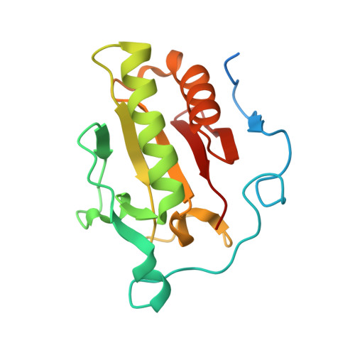
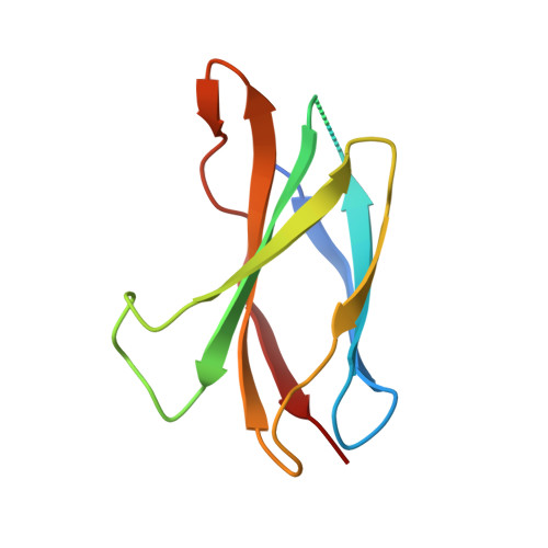
The mode of Hedgehog binding to Ihog homologues is not conserved across different phyla.

McLellan, J.S.Zheng, X.Hauk, G.Ghirlando, R.Beachy, P.A.Leahy, D.J.

(2008) Nature 455: 979-983

  • DOI: https://doi.org/10.1038/nature07358
  • Primary Citation of Related Structures:  
    3D1M

  • PubMed Abstract: 

    Hedgehog (Hh) proteins specify tissue pattern in metazoan embryos by forming gradients that emanate from discrete sites of expression and elicit concentration-dependent cellular differentiation or proliferation responses. Cellular responses to Hh and the movement of Hh through tissues are both precisely regulated, and abnormal Hh signalling has been implicated in human birth defects and cancer. Hh signalling is mediated by its amino-terminal domain (HhN), which is dually lipidated and secreted as part of a multivalent lipoprotein particle. Reception of the HhN signal is modulated by several cell-surface proteins on responding cells, including Patched (Ptc), Smoothened (Smo), Ihog (known as CDO or CDON in mammals) and the vertebrate-specific proteins Hip (also known as Hhip) and Gas1 (ref. 11). Drosophila Ihog and its vertebrate homologues CDO and BOC contain multiple immunoglobulin and fibronectin type III (FNIII) repeats, and the first FNIII repeat of Ihog binds Drosophila HhN in a heparin-dependent manner. Surprisingly, pull-down experiments suggest that a mammalian Sonic hedgehog N-terminal domain (ShhN) binds a non-orthologous FNIII repeat of CDO. Here we report biochemical, biophysical and X-ray structural studies of a complex between ShhN and the third FNIII repeat of CDO. We show that the ShhN-CDO interaction is completely unlike the HhN-Ihog interaction and requires calcium, which binds at a previously undetected site on ShhN. This site is conserved in nearly all Hh proteins and is a hotspot for mediating interactions between ShhN and CDO, Ptc, Hip and Gas1. Mutations in vertebrate Hh proteins causing holoprosencephaly and brachydactyly type A1 map to this calcium-binding site and disrupt interactions with these partners.


  • Organizational Affiliation

    Department of Biophysics and Biophysical Chemistry, Johns Hopkins University School of Medicine, Baltimore, Maryland 21205, USA.


Macromolecules
Find similar proteins by:  (by identity cutoff)  |  3D Structure
Entity ID: 1
MoleculeChains Sequence LengthOrganismDetailsImage
Sonic hedgehog protein
A, B
168Mus musculusMutation(s): 0 
Gene Names: ShhHhg1
UniProt & NIH Common Fund Data Resources
Find proteins for Q62226 (Mus musculus)
Explore Q62226 
Go to UniProtKB:  Q62226
IMPC:  MGI:98297
Entity Groups  
Sequence Clusters30% Identity50% Identity70% Identity90% Identity95% Identity100% Identity
UniProt GroupQ62226
Sequence Annotations
Expand
  • Reference Sequence
Find similar proteins by:  (by identity cutoff)  |  3D Structure
Entity ID: 2
MoleculeChains Sequence LengthOrganismDetailsImage
Cell adhesion molecule
C, D
102Homo sapiensMutation(s): 0 
Gene Names: CDONCDO
UniProt & NIH Common Fund Data Resources
Find proteins for Q4KMG0 (Homo sapiens)
Explore Q4KMG0 
Go to UniProtKB:  Q4KMG0
PHAROS:  Q4KMG0
GTEx:  ENSG00000064309 
Entity Groups  
Sequence Clusters30% Identity50% Identity70% Identity90% Identity95% Identity100% Identity
UniProt GroupQ4KMG0
Sequence Annotations
Expand
  • Reference Sequence
Experimental Data & Validation

Experimental Data

  • Method: X-RAY DIFFRACTION
  • Resolution: 1.70 Å
  • R-Value Free: 0.229 
  • R-Value Work: 0.186 
  • R-Value Observed: 0.189 
  • Space Group: C 2 2 21
Unit Cell:
Length ( Å )Angle ( ˚ )
a = 71.179α = 90
b = 98.582β = 90
c = 144.047γ = 90
Software Package:
Software NamePurpose
DENZOdata reduction
SCALEPACKdata scaling
MOLREPphasing
REFMACrefinement
PDB_EXTRACTdata extraction

Structure Validation

View Full Validation Report


Entry History 

Deposition Data

Revision History  (Full details and data files)

  • Version 1.0: 2008-09-23
    Type: Initial release
  • Version 1.1: 2011-07-13
    Changes: Advisory, Refinement description, Version format compliance
  • Version 1.2: 2017-10-25
    Changes: Refinement description
  • Version 1.3: 2024-02-21
    Changes: Data collection, Database references, Derived calculations