


Crystal structure of CYP2R1 in complex with vitamin D2.
Strushkevich, N.V., Tempel, W., Gilep, A.A., Loppnau, P., Arrowsmith, C.H., Edwards, A.M., Bountra, C., Wilkstrom, M., Bochkarev, A., Park, H.To be published.
Experimental Data Snapshot
Entity ID: 1 | |||||
|---|---|---|---|---|---|
| Molecule | Chains | Sequence Length | Organism | Details | Image |
| Cytochrome P450 2R1 | 481 | Homo sapiens | Mutation(s): 0 Gene Names: CYP2R1 EC: 1.14.14 Membrane Entity: Yes | 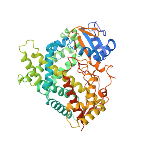 | |
UniProt & NIH Common Fund Data Resources | |||||
Find proteins for Q6VVX0 (Homo sapiens) Explore Q6VVX0 Go to UniProtKB: Q6VVX0 | |||||
PHAROS: Q6VVX0 GTEx: ENSG00000186104 | |||||
Entity Groups | |||||
| Sequence Clusters | 30% Identity50% Identity70% Identity90% Identity95% Identity100% Identity | ||||
| UniProt Group | Q6VVX0 | ||||
Sequence AnnotationsExpand | |||||
| |||||
| Ligands 3 Unique | |||||
|---|---|---|---|---|---|
| ID | Chains | Name / Formula / InChI Key | 2D Diagram | 3D Interactions | |
| HEM Query on HEM | BA [auth B], O [auth A] | PROTOPORPHYRIN IX CONTAINING FE C34 H32 Fe N4 O4 KABFMIBPWCXCRK-RGGAHWMASA-L |  | ||
| D2V Query on D2V | CA [auth B], P [auth A] | (3S,5Z,7E,22E)-9,10-secoergosta-5,7,10,22-tetraen-3-ol C28 H44 O MECHNRXZTMCUDQ-RKHKHRCZSA-N |  | ||
| UNX Query on UNX | AA [auth B] E [auth A] F [auth A] G [auth A] H [auth A] | UNKNOWN ATOM OR ION X |  | ||
Entity ID: 2 | |||||
|---|---|---|---|---|---|
| ID | Chains | Name | Type/Class | 2D Diagram | 3D Interactions |
| PRD_900012 Query on PRD_900012 | C, D | beta-cyclodextrin | Oligosaccharide / Drug delivery |  | |
| Length ( Å ) | Angle ( ˚ ) |
|---|---|
| a = 137.361 | α = 90 |
| b = 163.393 | β = 90 |
| c = 152.289 | γ = 90 |
| Software Name | Purpose |
|---|---|
| DENZO | data reduction |
| SCALEPACK | data scaling |
| REFMAC | refinement |
| PDB_EXTRACT | data extraction |
| ADSC | data collection |
| HKL-3000 | data reduction |
| REFMAC | phasing |
RCSB PDB (citation) is hosted by
RCSB PDB is a member of the


