


3CXD
Crystal structure of anti-osteopontin antibody 23C3 in complex with its epitope peptide
- PDB DOI: https://doi.org/10.2210/pdb3CXD/pdb
- Classification: IMMUNE SYSTEM
- Organism(s): Mus musculus, Homo sapiens
- Mutation(s): No
- Deposited: 2008-04-24 Released: 2008-10-14
Experimental Data Snapshot
- Method: X-RAY DIFFRACTION
- Resolution: 2.80 Å
- R-Value Free: 0.292
- R-Value Work: 0.248
- R-Value Observed: 0.251
wwPDB Validation 3D Report Full Report

This is version 2.1 of the entry. See complete history.

Macromolecules
Find similar proteins by:
(by identity cutoff) | 3D Structure
Entity ID: 1 | |||||
|---|---|---|---|---|---|
| Molecule | Chains | Sequence Length | Organism | Details | Image |
| Fab fragment of anti-osteopontin antibody 23C3, Light chain | A [auth L] | 213 | Mus musculus | Mutation(s): 0 | 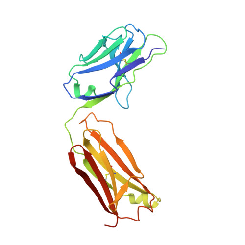 |
Entity Groups | |||||
| Sequence Clusters | 30% Identity50% Identity70% Identity90% Identity95% Identity100% Identity | ||||
Sequence AnnotationsExpand | |||||
| |||||
Find similar proteins by:
(by identity cutoff) | 3D Structure
Entity ID: 2 | |||||
|---|---|---|---|---|---|
| Molecule | Chains | Sequence Length | Organism | Details | Image |
| Fab fragment of anti-osteopontin antibody 23C3, Heavy chain | B [auth H] | 216 | Mus musculus | Mutation(s): 0 | 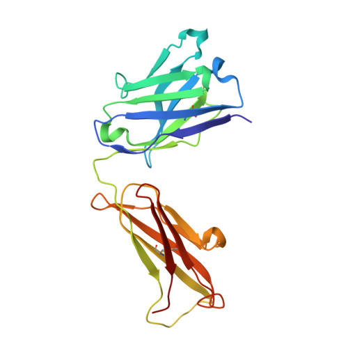 |
Entity Groups | |||||
| Sequence Clusters | 30% Identity50% Identity70% Identity90% Identity95% Identity100% Identity | ||||
Sequence AnnotationsExpand | |||||
| |||||
Find similar proteins by: Sequence | 3D Structure
Entity ID: 3 | |||||
|---|---|---|---|---|---|
| Molecule | Chains | Sequence Length | Organism | Details | Image |
| a peptide from osteopontin | C [auth P] | 12 | N/A | Mutation(s): 0 | 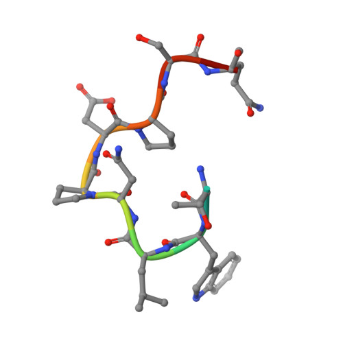 |
UniProt & NIH Common Fund Data Resources | |||||
Find proteins for P10451 (Homo sapiens) Explore P10451 Go to UniProtKB: P10451 | |||||
PHAROS: P10451 GTEx: ENSG00000118785 | |||||
Entity Groups | |||||
| Sequence Clusters | 30% Identity50% Identity70% Identity90% Identity95% Identity100% Identity | ||||
| UniProt Group | P10451 | ||||
Sequence AnnotationsExpand | |||||
| |||||
Oligosaccharides
Entity ID: 4 | |||||
|---|---|---|---|---|---|
| Molecule | Chains | Length | 2D Diagram | Glycosylation | 3D Interactions |
| alpha-D-mannopyranose-(1-3)-beta-D-mannopyranose-(1-4)-2-acetamido-2-deoxy-beta-D-glucopyranose-(1-4)-2-acetamido-2-deoxy-beta-D-glucopyranose | D [auth A] | 4 |  | N-Glycosylation | |
Glycosylation Resources | |||||
GlyTouCan: G81315DD GlyCosmos: G81315DD GlyGen: G81315DD | |||||
Experimental Data & Validation
Experimental Data
- Method: X-RAY DIFFRACTION
- Resolution: 2.80 Å
- R-Value Free: 0.292
- R-Value Work: 0.248
- R-Value Observed: 0.251
- Space Group: P 21 21 21
Unit Cell:
| Length ( Å ) | Angle ( ˚ ) |
|---|---|
| a = 42.318 | α = 90 |
| b = 92.02 | β = 90 |
| c = 122.483 | γ = 90 |
| Software Name | Purpose |
|---|---|
| d*TREK | data scaling |
| PHASER | phasing |
| REFMAC | refinement |
| PDB_EXTRACT | data extraction |
| CrystalClear | data collection |
| CrystalClear | data reduction |
| CrystalClear | data scaling |
Entry History
Deposition Data
Revision History (Full details and data files)
- Version 1.0: 2008-10-14
Type: Initial release - Version 1.1: 2011-07-13
Changes: Non-polymer description, Version format compliance - Version 2.0: 2020-07-29
Type: Remediation
Reason: Carbohydrate remediation
Changes: Atomic model, Data collection, Derived calculations, Structure summary - Version 2.1: 2023-11-01
Changes: Data collection, Database references, Refinement description, Structure summary
