3CUS

Structure of a double ILE/PHE mutant of NI-FE hydrogenase refined at 2.2 angstrom resolution


Experimental Data Snapshot

  • Method: X-RAY DIFFRACTION
  • Resolution: 2.20 Å
  • R-Value Free: 0.222 
  • R-Value Work: 0.185 
  • R-Value Observed: 0.187 

wwPDB Validation   3D Report Full Report


This is version 1.4 of the entry. See complete history


Literature

Experimental approaches to kinetics of gas diffusion in hydrogenase

Leroux, F.Dementin, S.Burlat, B.Cournac, L.Volbeda, A.Champ, S.Martin, L.Guigliarelli, B.Bertrand, P.Fontecilla-Camps, J.Rousset, M.

(2008) Proc Natl Acad Sci U S A 105: 11188-11193

  • DOI: https://doi.org/10.1073/pnas.0803689105
  • Primary Citation of Related Structures:  
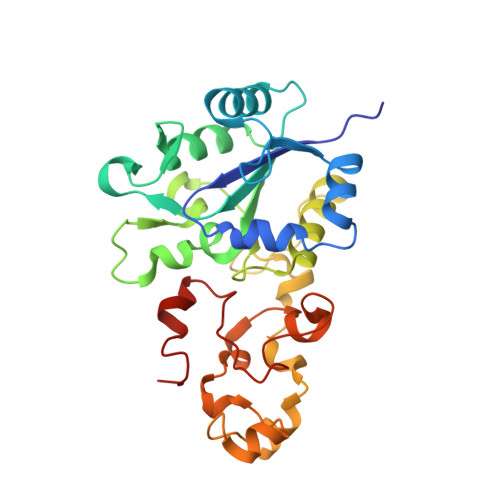
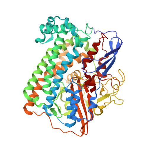
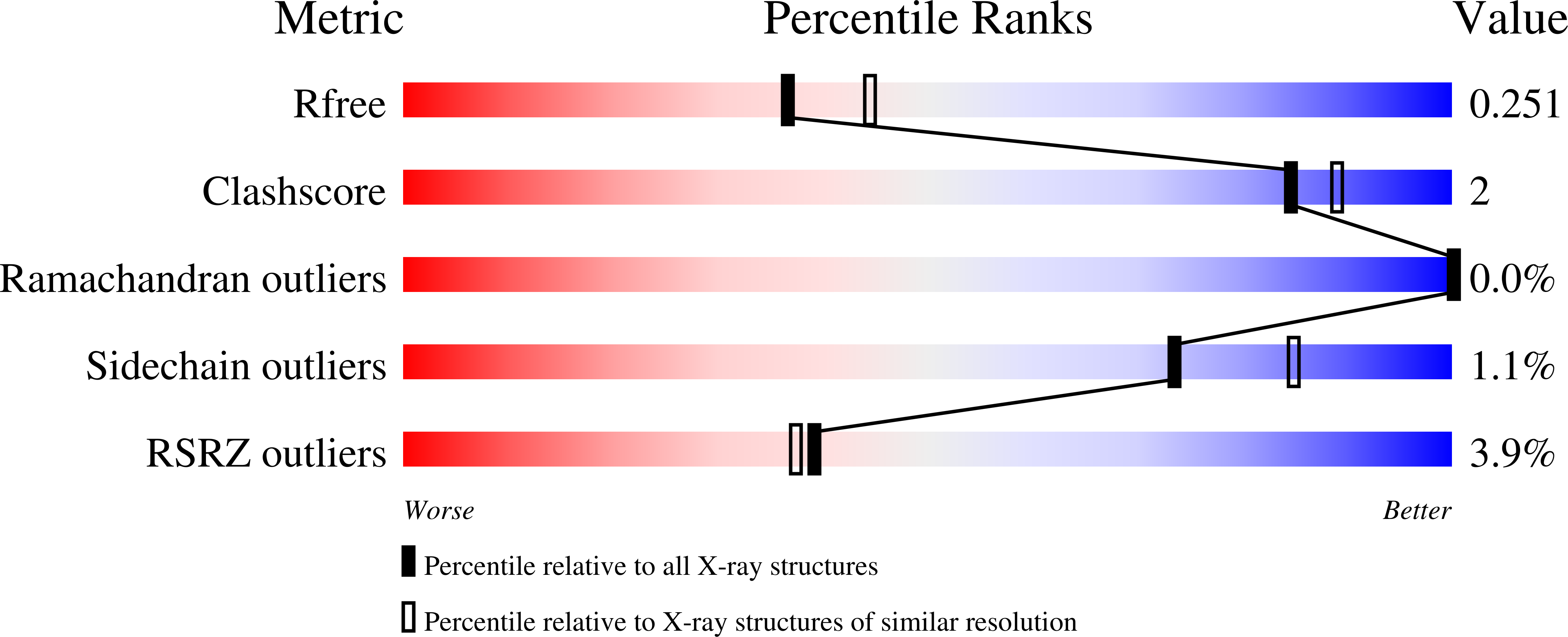
    3CUR, 3CUS

  • PubMed Abstract: 

    Hydrogenases, which catalyze H(2) to H(+) conversion as part of the bioenergetic metabolism of many microorganisms, are among the metalloenzymes for which a gas-substrate tunnel has been described by using crystallography and molecular dynamics. However, the correlation between protein structure and gas-diffusion kinetics is unexplored. Here, we introduce two quantitative methods for probing the rates of diffusion within hydrogenases. One uses protein film voltammetry to resolve the kinetics of binding and release of the competitive inhibitor CO; the other is based on interpreting the yield in the isotope exchange assay. We study structurally characterized mutants of a NiFe hydrogenase, and we show that two mutations, which significantly narrow the tunnel near the entrance of the catalytic center, decrease the rates of diffusion of CO and H(2) toward and from the active site by up to 2 orders of magnitude. This proves the existence of a functional channel, which matches the hydrophobic cavity found in the crystal. However, the changes in diffusion rates do not fully correlate with the obstruction induced by the mutation and deduced from the x-ray structures. Our results demonstrate the necessity of measuring diffusion rates and emphasize the role of side-chain dynamics in determining these.


  • Organizational Affiliation

    Centre National de la Recherche Scientifique, Institut de Biologie Structurale et Microbiologie, Unité Propre de Recherche 9036, Unité de Bioénergétique et Ingénierie des Protéines, Marseille, France.


Macromolecules
Find similar proteins by:  (by identity cutoff)  |  3D Structure
Entity ID: 1
MoleculeChains Sequence LengthOrganismDetailsImage
Periplasmic [NiFe] hydrogenase small subunitA,
C [auth B],
E [auth C]
264Solidesulfovibrio fructosivoransMutation(s): 0 
Gene Names: hydA
EC: 1.12.2.1
UniProt
Find proteins for P18187 (Solidesulfovibrio fructosivorans)
Explore P18187 
Go to UniProtKB:  P18187
Entity Groups  
Sequence Clusters30% Identity50% Identity70% Identity90% Identity95% Identity100% Identity
UniProt GroupP18187
Sequence Annotations
Expand
  • Reference Sequence
Find similar proteins by:  (by identity cutoff)  |  3D Structure
Entity ID: 2
MoleculeChains Sequence LengthOrganismDetailsImage
Periplasmic [NiFe] hydrogenase large subunitB [auth Q],
D [auth R],
F [auth S]
549Solidesulfovibrio fructosivoransMutation(s): 2 
Gene Names: hydB
EC: 1.12.2.1
UniProt
Find proteins for P18188 (Solidesulfovibrio fructosivorans)
Explore P18188 
Go to UniProtKB:  P18188
Entity Groups  
Sequence Clusters30% Identity50% Identity70% Identity90% Identity95% Identity100% Identity
UniProt GroupP18188
Sequence Annotations
Expand
  • Reference Sequence
Small Molecules
Ligands 6 Unique
IDChains Name / Formula / InChI Key2D Diagram3D Interactions
SF4
Query on SF4

Download Ideal Coordinates CCD File 
G [auth A]
I [auth A]
P [auth B]
R [auth B]
W [auth C]
G [auth A],
I [auth A],
P [auth B],
R [auth B],
W [auth C],
Y [auth C]
IRON/SULFUR CLUSTER
Fe4 S4
LJBDFODJNLIPKO-UHFFFAOYSA-N
F3S
Query on F3S

Download Ideal Coordinates CCD File 
H [auth A],
Q [auth B],
X [auth C]
FE3-S4 CLUSTER
Fe3 S4
FCXHZBQOKRZXKS-UHFFFAOYSA-N
FCO
Query on FCO

Download Ideal Coordinates CCD File 
BA [auth S],
M [auth Q],
U [auth R]
CARBONMONOXIDE-(DICYANO) IRON
C3 Fe N2 O
VBQUCMTXYFMTTE-UHFFFAOYSA-N
GOL
Query on GOL

Download Ideal Coordinates CCD File 
CA [auth S],
J [auth A],
N [auth Q],
O [auth Q],
V [auth R]
GLYCEROL
C3 H8 O3
PEDCQBHIVMGVHV-UHFFFAOYSA-N
NI
Query on NI

Download Ideal Coordinates CCD File 
K [auth Q],
S [auth R],
Z [auth S]
NICKEL (II) ION
Ni
VEQPNABPJHWNSG-UHFFFAOYSA-N
MG
Query on MG

Download Ideal Coordinates CCD File 
AA [auth S],
L [auth Q],
T [auth R]
MAGNESIUM ION
Mg
JLVVSXFLKOJNIY-UHFFFAOYSA-N
Modified Residues  1 Unique
IDChains TypeFormula2D DiagramParent
CSO
Query on CSO
B [auth Q],
D [auth R],
F [auth S]
L-PEPTIDE LINKINGC3 H7 N O3 SCYS
Experimental Data & Validation

Experimental Data

  • Method: X-RAY DIFFRACTION
  • Resolution: 2.20 Å
  • R-Value Free: 0.222 
  • R-Value Work: 0.185 
  • R-Value Observed: 0.187 
  • Space Group: P 1 21 1
Unit Cell:
Length ( Å )Angle ( ˚ )
a = 62.4α = 90
b = 99.96β = 92.23
c = 182.69γ = 90
Software Package:
Software NamePurpose
AMoREphasing
REFMACrefinement
ADSCdata collection
XDSdata reduction
XDSdata scaling

Structure Validation

View Full Validation Report


Entry History 

Deposition Data

  • Released Date: 2008-08-05 
  • Deposition Author(s): Volbeda, A.

Revision History  (Full details and data files)

  • Version 1.0: 2008-08-05
    Type: Initial release
  • Version 1.1: 2011-07-13
    Changes: Non-polymer description, Version format compliance
  • Version 1.2: 2011-09-07
    Changes: Database references
  • Version 1.3: 2023-11-01
    Changes: Data collection, Database references, Derived calculations, Refinement description
  • Version 1.4: 2023-11-15
    Changes: Data collection