3CU0

human beta 1,3-glucuronyltransferase I (GlcAT-I) in complex with UDP and GAL-GAL(6-SO4)-XYL(2-PO4)-O-SER


Experimental Data Snapshot

  • Method: X-RAY DIFFRACTION
  • Resolution: 1.90 Å
  • R-Value Free: 0.234 
  • R-Value Work: 0.210 
  • R-Value Observed: 0.210 

wwPDB Validation   3D Report Full Report


Ligand Structure Quality Assessment 


This is version 2.1 of the entry. See complete history


Literature

2-o-phosphorylation of xylose and 6-o-sulfation of galactose in the protein linkage region of glycosaminoglycans influence the glucuronyltransferase-I activity involved in the linkage region synthesis.

Tone, Y.Pedersen, L.C.Yamamoto, T.Izumikawa, T.Kitagawa, H.Nishihara, J.Tamura, J.Negishi, M.Sugahara, K.

(2008) J Biol Chem 283: 16801-16807

  • DOI: https://doi.org/10.1074/jbc.M709556200
  • Primary Citation of Related Structures:  
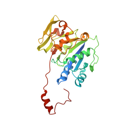
    3CU0

  • PubMed Abstract: 

    Sulfated glycosaminoglycans (GAGs), including heparan sulfate and chondroitin sulfate, are synthesized on the so-called common GAG-protein linkage region (GlcUAbeta1-3Galbeta1-3Galbeta1-4Xylbeta1-O-Ser) of core proteins, which is formed by the stepwise addition of monosaccharide residues by the respective specific glycosyltransferases. Glucuronyltransferase-I (GlcAT-I) is the key enzyme that completes the synthesis of this linkage region, which is a prerequisite for the conversion of core proteins to functional proteoglycans bearing GAGs. The Xyl and Gal residues in the linkage region can be modified by phosphorylation and sulfation, respectively, although the biological significance of these modifications remains to be clarified. Here we present evidence that these modifications can significantly influence the catalytic activity of GlcAT-I. Enzyme assays showed that the synthetic substrates, Gal-Gal-Xyl(2-O-phosphate)-O-Ser and Gal-Gal(6-O-sulfate)-Xyl(2-O-phosphate)-O-Ser, served as better substrates than the unmodified compound, whereas Gal(6-O-sulfate)-Gal-Xyl(2-O-phosphate)-O-Ser exhibited no acceptor activity. The crystal structure of the catalytic domain of GlcAT-I with UDP and Gal-Gal(6-O-sulfate)-Xyl(2-O-phosphate)-O-Ser bound revealed that the Xyl(2-O-phosphate)-O-Ser is disordered and the 6-O-sulfate forms interactions with Gln(318) from the second GlcAT-I monomer in the dimeric enzyme. The results indicate the possible involvement of these modifications in the processing and maturation of the growing linkage region oligosaccharide required for the assembly of GAG chains.


  • Organizational Affiliation

    Department of Biochemistry, Kobe Pharmaceutical University, Higashinada-ku, Kobe 658-8558, Japan.


Macromolecules
Find similar proteins by:  (by identity cutoff)  |  3D Structure
Entity ID: 1
MoleculeChains Sequence LengthOrganismDetailsImage
Galactosylgalactosylxylosylprotein 3-beta-glucuronosyltransferase 3
A, B
281Homo sapiensMutation(s): 0 
Gene Names: B3GAT3
EC: 2.4.1.135
UniProt & NIH Common Fund Data Resources
Find proteins for O94766 (Homo sapiens)
Explore O94766 
Go to UniProtKB:  O94766
PHAROS:  O94766
GTEx:  ENSG00000149541 
Entity Groups  
Sequence Clusters30% Identity50% Identity70% Identity90% Identity95% Identity100% Identity
UniProt GroupO94766
Sequence Annotations
Expand
  • Reference Sequence
Oligosaccharides

Help

Entity ID: 2
MoleculeChains Length2D Diagram Glycosylation3D Interactions
beta-D-galactopyranose-(1-3)-beta-D-galactopyranose
C, D
2N/A
Glycosylation Resources
GlyTouCan:  G95894VW
GlyCosmos:  G95894VW
GlyGen:  G95894VW
Experimental Data & Validation

Experimental Data

  • Method: X-RAY DIFFRACTION
  • Resolution: 1.90 Å
  • R-Value Free: 0.234 
  • R-Value Work: 0.210 
  • R-Value Observed: 0.210 
  • Space Group: P 1 21 1
Unit Cell:
Length ( Å )Angle ( ˚ )
a = 57.047α = 90
b = 48.399β = 92.4
c = 103.708γ = 90
Software Package:
Software NamePurpose
CNSrefinement
StructureStudiodata collection
DENZOdata reduction
SCALEPACKdata scaling
CNSphasing

Structure Validation

View Full Validation Report


Ligand Structure Quality Assessment 


Entry History 

Deposition Data

Revision History  (Full details and data files)

  • Version 1.0: 2008-05-06
    Type: Initial release
  • Version 1.1: 2011-07-13
    Changes: Version format compliance
  • Version 2.0: 2020-07-29
    Type: Remediation
    Reason: Carbohydrate remediation
    Changes: Atomic model, Data collection, Database references, Derived calculations, Structure summary
  • Version 2.1: 2023-08-30
    Changes: Data collection, Database references, Refinement description, Structure summary