Structural basis for substrate specificity in phosphate binding (beta/alpha)8-barrels: D-allulose 6-phosphate 3-epimerase from Escherichia coli K-12.
Chan, K.K., Fedorov, A.A., Fedorov, E.V., Almo, S.C., Gerlt, J.A.(2008) Biochemistry 47: 9608-9617
- PubMed: 18700786
- DOI: https://doi.org/10.1021/bi800821v
- Primary Citation of Related Structures:
3CT7, 3CTL - PubMed Abstract:
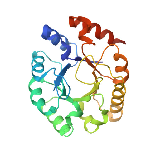
Enzymes that share the (beta/alpha) 8-barrel fold catalyze a diverse range of reactions. Many utilize phosphorylated substrates and share a conserved C-terminal (beta/alpha) 2-quarter barrel subdomain that provides a binding motif for the dianionic phosphate group. We recently reported functional and structural studies of d-ribulose 5-phosphate 3-epimerase (RPE) from Streptococcus pyogenes that catalyzes the equilibration of the pentulose 5-phosphates d-ribulose 5-phosphate and d-xylulose 5-phosphate in the pentose phosphate pathway [J. Akana, A. A. Fedorov, E. Fedorov, W. R. P. Novack, P. C. Babbitt, S. C. Almo, and J. A. Gerlt (2006) Biochemistry 45, 2493-2503]. We now report functional and structural studies of d-allulose 6-phosphate 3-epimerase (ALSE) from Escherichia coli K-12 that catalyzes the equilibration of the hexulose 6-phosphates d-allulose 6-phosphate and d-fructose 6-phosphate in a catabolic pathway for d-allose. ALSE and RPE prefer their physiological substrates but are promiscuous for each other's substrate. The active sites (RPE complexed with d-xylitol 5-phosphate and ALSE complexed with d-glucitol 6-phosphate) are superimposable (as expected from their 39% sequence identity), with the exception of the phosphate binding motif. The loop following the eighth beta-strand in ALSE is one residue longer than the homologous loop in RPE, so the binding site for the hexulose 6-phosphate substrate/product in ALSE is elongated relative to that for the pentulose 5-phosphate substrate/product in RPE. We constructed three single-residue deletion mutants of the loop in ALSE, DeltaT196, DeltaS197 and DeltaG198, to investigate the structural bases for the differing substrate specificities; for each, the promiscuity is altered so that d-ribulose 5-phosphate is the preferred substrate. The changes in k cat/ K m are dominated by changes in k cat, suggesting that substrate discrimination results from differential transition state stabilization. In both ALSE and RPE, the phosphate group hydrogen bonds not only with the conserved motif but also with an active site loop following the sixth beta-strand, providing a potential structural mechanism for coupling substrate binding with catalysis.
Organizational Affiliation:
Departments of Biochemistry and Chemistry, University of Illinois at Urbana-Champaign, 600 South Mathews Avenue, Urbana, Illinois 61801, USA.