3CSU

CATALYTIC TRIMER OF ESCHERICHIA COLI ASPARTATE TRANSCARBAMOYLASE


Experimental Data Snapshot

  • Method: X-RAY DIFFRACTION
  • Resolution: 1.88 Å
  • R-Value Free: 0.291 
  • R-Value Work: 0.190 

wwPDB Validation   3D Report Full Report


This is version 1.3 of the entry. See complete history


Literature

Assessment of the allosteric mechanism of aspartate transcarbamoylase based on the crystalline structure of the unregulated catalytic subunit.

Beernink, P.T.Endrizzi, J.A.Alber, T.Schachman, H.K.

(1999) Proc Natl Acad Sci U S A 96: 5388-5393

  • DOI: https://doi.org/10.1073/pnas.96.10.5388
  • Primary Citation of Related Structures:  
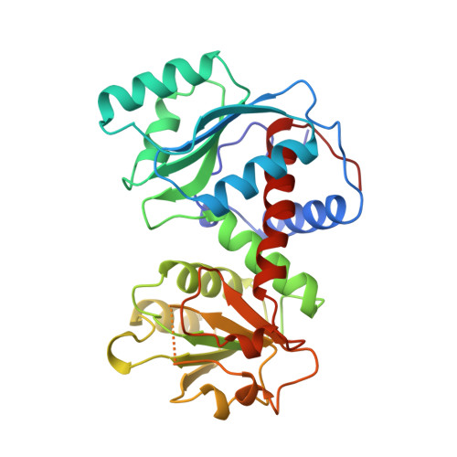
    3CSU

  • PubMed Abstract: 

    The lack of knowledge of the three-dimensional structure of the trimeric, catalytic (C) subunit of aspartate transcarbamoylase (ATCase) has impeded understanding of the allosteric regulation of this enzyme and left unresolved the mechanism by which the active, unregulated C trimers are inactivated on incorporation into the unliganded (taut or T state) holoenzyme. Surprisingly, the isolated C trimer, based on the 1.9-A crystal structure reported here, resembles more closely the trimers in the T state enzyme than in the holoenzyme:bisubstrate-analog complex, which has been considered as the active, relaxed (R) state enzyme. Unlike the C trimer in either the T state or bisubstrate-analog-bound holoenzyme, the isolated C trimer lacks 3-fold symmetry, and the active sites are partially disordered. The flexibility of the C trimer, contrasted to the highly constrained T state ATCase, suggests that regulation of the holoenzyme involves modulating the potential for conformational changes essential for catalysis. Large differences in structure between the active C trimer and the holoenzyme:bisubstrate-analog complex call into question the view that this complex represents the activated R state of ATCase.


  • Organizational Affiliation

    Department of Molecular and Cell Biology and Virus Laboratory, University of California, Berkeley, CA 94720-3206, USA.


Macromolecules
Find similar proteins by:  (by identity cutoff)  |  3D Structure
Entity ID: 1
MoleculeChains Sequence LengthOrganismDetailsImage
PROTEIN (ASPARTATE CARBAMOYLTRANSFERASE)
A, B, C
310Escherichia coli K-12Mutation(s): 0 
Gene Names: PYRB
EC: 2.1.3.2
UniProt
Find proteins for P0A786 (Escherichia coli (strain K12))
Explore P0A786 
Go to UniProtKB:  P0A786
Entity Groups  
Sequence Clusters30% Identity50% Identity70% Identity90% Identity95% Identity100% Identity
UniProt GroupP0A786
Sequence Annotations
Expand
  • Reference Sequence
Small Molecules
Ligands 1 Unique
IDChains Name / Formula / InChI Key2D Diagram3D Interactions
CA
Query on CA

Download Ideal Coordinates CCD File 
D [auth B],
E [auth C]
CALCIUM ION
Ca
BHPQYMZQTOCNFJ-UHFFFAOYSA-N
Experimental Data & Validation

Experimental Data

  • Method: X-RAY DIFFRACTION
  • Resolution: 1.88 Å
  • R-Value Free: 0.291 
  • R-Value Work: 0.190 
  • Space Group: P 21 21 21
Unit Cell:
Length ( Å )Angle ( ˚ )
a = 56.12α = 90
b = 82.54β = 90
c = 210.52γ = 90
Software Package:
Software NamePurpose
AMoREphasing
TNTrefinement
MOSFLMdata reduction
CCP4data scaling

Structure Validation

View Full Validation Report


Entry History 

Deposition Data

Revision History  (Full details and data files)

  • Version 1.0: 1999-05-11
    Type: Initial release
  • Version 1.1: 2008-04-26
    Changes: Version format compliance
  • Version 1.2: 2011-07-13
    Changes: Source and taxonomy, Version format compliance
  • Version 1.3: 2023-08-30
    Changes: Data collection, Database references, Derived calculations, Refinement description