


Crystal structure of p38delta kinase.
Atwell, S., Burley, S.K., Houle, A., Ramos, A., Sauder, J.M.To be published.
Experimental Data Snapshot
wwPDB Validation 3D Report Full Report

Entity ID: 1 | |||||
|---|---|---|---|---|---|
| Molecule | Chains | Sequence Length | Organism | Details | Image |
| Mitogen-activated protein kinase 13 | 353 | Homo sapiens | Mutation(s): 2 Gene Names: MAPK13, PRKM13, SAPK4 EC: 2.7.11.24 | 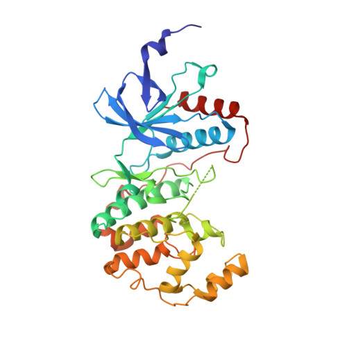 | |
UniProt & NIH Common Fund Data Resources | |||||
Find proteins for O15264 (Homo sapiens) Explore O15264 Go to UniProtKB: O15264 | |||||
PHAROS: O15264 GTEx: ENSG00000156711 | |||||
Entity Groups | |||||
| Sequence Clusters | 30% Identity50% Identity70% Identity90% Identity95% Identity100% Identity | ||||
| UniProt Group | O15264 | ||||
Sequence AnnotationsExpand | |||||
| |||||
| Length ( Å ) | Angle ( ˚ ) |
|---|---|
| a = 66.271 | α = 90 |
| b = 71.305 | β = 90 |
| c = 97.933 | γ = 90 |
| Software Name | Purpose |
|---|---|
| MAR345 | data collection |
| EPMR | phasing |
| REFMAC | refinement |
| MOSFLM | data reduction |
| SCALA | data scaling |
RCSB PDB (citation) is hosted by
RCSB PDB is a member of the

