


X-ray crystal structure of NUDIX hydrolase from Nitrosomonas europaea.
Osipiuk, J., Xu, X., Zheng, H., Savchenko, A., Edwards, A.M., Joachimiak, A.To be published.
Experimental Data Snapshot
wwPDB Validation 3D Report Full Report

Entity ID: 1 | |||||
|---|---|---|---|---|---|
| Molecule | Chains | Sequence Length | Organism | Details | Image |
| NUDIX hydrolase | 189 | Nitrosomonas europaea ATCC 19718 | Mutation(s): 0 Gene Names: NE1158 | 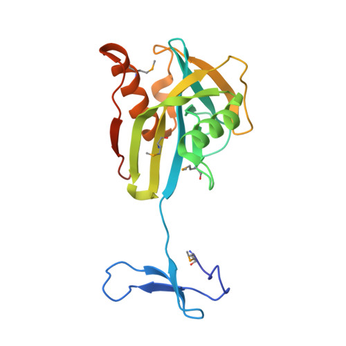 | |
UniProt | |||||
Find proteins for Q82VD6 (Nitrosomonas europaea (strain ATCC 19718 / CIP 103999 / KCTC 2705 / NBRC 14298)) Explore Q82VD6 Go to UniProtKB: Q82VD6 | |||||
Entity Groups | |||||
| Sequence Clusters | 30% Identity50% Identity70% Identity90% Identity95% Identity100% Identity | ||||
| UniProt Group | Q82VD6 | ||||
Sequence AnnotationsExpand | |||||
| |||||
| Ligands 5 Unique | |||||
|---|---|---|---|---|---|
| ID | Chains | Name / Formula / InChI Key | 2D Diagram | 3D Interactions | |
| GOL Query on GOL | K [auth A], P [auth B], T [auth C], X [auth D] | GLYCEROL C3 H8 O3 PEDCQBHIVMGVHV-UHFFFAOYSA-N |  | ||
| ZN Query on ZN | E [auth A], L [auth B], Q [auth C], U [auth D] | ZINC ION Zn PTFCDOFLOPIGGS-UHFFFAOYSA-N |  | ||
| EDO Query on EDO | J [auth A], S [auth C], W [auth D] | 1,2-ETHANEDIOL C2 H6 O2 LYCAIKOWRPUZTN-UHFFFAOYSA-N |  | ||
| CL Query on CL | G [auth A] H [auth A] I [auth A] N [auth B] O [auth B] | CHLORIDE ION Cl VEXZGXHMUGYJMC-UHFFFAOYSA-M |  | ||
| NA Query on NA | F [auth A], M [auth B] | SODIUM ION Na FKNQFGJONOIPTF-UHFFFAOYSA-N |  | ||
| Modified Residues 1 Unique | |||||
|---|---|---|---|---|---|
| ID | Chains | Type | Formula | 2D Diagram | Parent |
| MSE Query on MSE | A, B, C, D | L-PEPTIDE LINKING | C5 H11 N O2 Se |  | MET |
| Length ( Å ) | Angle ( ˚ ) |
|---|---|
| a = 174.781 | α = 90 |
| b = 49.506 | β = 106.28 |
| c = 110.19 | γ = 90 |
| Software Name | Purpose |
|---|---|
| DENZO | data reduction |
| SCALEPACK | data scaling |
| REFMAC | refinement |
| PDB_EXTRACT | data extraction |
| SBC-Collect | data collection |
| HKL-3000 | data reduction |
| SHELXD | phasing |
| MLPHARE | phasing |
| DM | phasing |
| SOLVE | phasing |
| RESOLVE | phasing |
| HKL-3000 | phasing |
RCSB PDB (citation) is hosted by
RCSB PDB is a member of the

