3CMM

Crystal Structure of the Uba1-Ubiquitin Complex


Experimental Data Snapshot

  • Method: X-RAY DIFFRACTION
  • Resolution: 2.70 Å
  • R-Value Free: 0.247 
  • R-Value Work: 0.191 
  • R-Value Observed: 0.194 

wwPDB Validation   3D Report Full Report


This is version 1.3 of the entry. See complete history


Literature

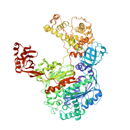
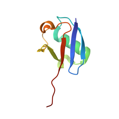
Structural insights into E1-catalyzed ubiquitin activation and transfer to conjugating enzymes.

Lee, I.Schindelin, H.

(2008) Cell 134: 268-278

  • DOI: https://doi.org/10.1016/j.cell.2008.05.046
  • Primary Citation of Related Structures:  
    3CMM

  • PubMed Abstract: 

    Ubiquitin (Ub) and ubiquitin-like proteins (Ubls) are conjugated to their targets by specific cascades involving three classes of enzymes, E1, E2, and E3. Each E1 adenylates the C terminus of its cognate Ubl, forms a E1 approximately Ubl thioester intermediate, and ultimately generates a thioester-linked E2 approximately Ubl product. We have determined the crystal structure of yeast Uba1, revealing a modular architecture with individual domains primarily mediating these specific activities. The negatively charged C-terminal ubiquitin-fold domain (UFD) is primed for binding of E2s and recognizes their positively charged first alpha helix via electrostatic interactions. In addition, a mobile loop from the domain harboring the E1 catalytic cysteine contributes to E2 binding. Significant, experimentally observed motions in the UFD around a hinge in the linker connecting this domain to the rest of the enzyme suggest a conformation-dependent mechanism for the transthioesterification function of Uba1; however, this mechanism clearly differs from that of other E1 enzymes.


  • Organizational Affiliation

    Department of Biochemistry and Cell Biology, Stony Brook University, Stony Brook, NY 11794-5215, USA.


Macromolecules
Find similar proteins by:  (by identity cutoff)  |  3D Structure
Entity ID: 1
MoleculeChains Sequence LengthOrganismDetailsImage
Ubiquitin-activating enzyme E1 1A,
B [auth C]
1,015Saccharomyces cerevisiaeMutation(s): 0 
Gene Names: UBA1
EC: 6.3.2.19
UniProt
Find proteins for P22515 (Saccharomyces cerevisiae (strain ATCC 204508 / S288c))
Explore P22515 
Go to UniProtKB:  P22515
Entity Groups  
Sequence Clusters30% Identity50% Identity70% Identity90% Identity95% Identity100% Identity
UniProt GroupP22515
Sequence Annotations
Expand
  • Reference Sequence
Find similar proteins by:  (by identity cutoff)  |  3D Structure
Entity ID: 2
MoleculeChains Sequence LengthOrganismDetailsImage
UbiquitinC [auth B],
D
76Saccharomyces cerevisiaeMutation(s): 0 
Gene Names: UBI1RPL40A
UniProt
Find proteins for P0CG63 (Saccharomyces cerevisiae (strain ATCC 204508 / S288c))
Explore P0CG63 
Go to UniProtKB:  P0CG63
Entity Groups  
Sequence Clusters30% Identity50% Identity70% Identity90% Identity95% Identity100% Identity
UniProt GroupP0CG63
Sequence Annotations
Expand
  • Reference Sequence
Experimental Data & Validation

Experimental Data

  • Method: X-RAY DIFFRACTION
  • Resolution: 2.70 Å
  • R-Value Free: 0.247 
  • R-Value Work: 0.191 
  • R-Value Observed: 0.194 
  • Space Group: P 21 21 21
Unit Cell:
Length ( Å )Angle ( ˚ )
a = 115.363α = 90
b = 118.564β = 90
c = 207.567γ = 90
Software Package:
Software NamePurpose
DENZOdata reduction
SCALEPACKdata scaling
REFMACrefinement
PDB_EXTRACTdata extraction
CBASSdata collection
PHASERphasing

Structure Validation

View Full Validation Report


Entry History 

Deposition Data

Revision History  (Full details and data files)

  • Version 1.0: 2008-08-05
    Type: Initial release
  • Version 1.1: 2011-07-13
    Changes: Advisory, Version format compliance
  • Version 1.2: 2017-10-25
    Changes: Refinement description
  • Version 1.3: 2024-02-21
    Changes: Data collection, Database references, Derived calculations, Refinement description