3CLC

Crystal Structure of the Restriction-Modification Controller Protein C.Esp1396I Tetramer in Complex with its Natural 35 Base-Pair Operator


Experimental Data Snapshot

  • Method: X-RAY DIFFRACTION
  • Resolution: 2.80 Å
  • R-Value Free: 0.239 
  • R-Value Work: 0.207 
  • R-Value Observed: 0.209 

wwPDB Validation   3D Report Full Report


This is version 1.2 of the entry. See complete history


Literature

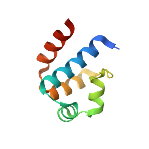
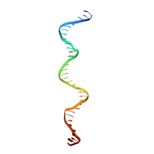
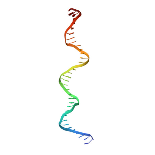
Structural analysis of the genetic switch that regulates the expression of restriction-modification genes.

McGeehan, J.E.Streeter, S.D.Thresh, S.J.Ball, N.Ravelli, R.B.Kneale, G.G.

(2008) Nucleic Acids Res 36: 4778-4787

  • DOI: https://doi.org/10.1093/nar/gkn448
  • Primary Citation of Related Structures:  
    3CLC

  • PubMed Abstract: 

    Controller (C) proteins regulate the timing of the expression of restriction and modification (R-M) genes through a combination of positive and negative feedback circuits. A single dimer bound to the operator switches on transcription of the C-gene and the endonuclease gene; at higher concentrations, a second dimer bound adjacently switches off these genes. Here we report the first structure of a C protein-DNA operator complex, consisting of two C protein dimers bound to the native 35 bp operator sequence of the R-M system Esp1396I. The structure reveals a role for both direct and indirect DNA sequence recognition. The structure of the DNA in the complex is highly distorted, with severe compression of the minor groove resulting in a 50 degrees bend within each operator site, together with a large expansion of the major groove in the centre of the DNA sequence. Cooperative binding between dimers governs the concentration-dependent activation-repression switch and arises, in part, from the interaction of Glu25 and Arg35 side chains at the dimer-dimer interface. Competition between Arg35 and an equivalent residue of the sigma(70) subunit of RNA polymerase for the Glu25 site underpins the switch from activation to repression of the endonuclease gene.


  • Organizational Affiliation

    Biophysics Laboratories, School of Biological Sciences, Institute of Biomedical and Biomolecular Sciences, University of Portsmouth, Portsmouth PO1 2DT, UK.


Macromolecules

Find similar proteins by:  (by identity cutoff)  |  3D Structure
Entity ID: 1
MoleculeChains Sequence LengthOrganismDetailsImage
Regulatory protein
A, B, C, D
82Enterobacter sp.Mutation(s): 0 
Gene Names: esp1396IC
UniProt
Find proteins for Q8GGH0 (Enterobacter sp. RFL1396)
Explore Q8GGH0 
Go to UniProtKB:  Q8GGH0
Entity Groups  
Sequence Clusters30% Identity50% Identity70% Identity90% Identity95% Identity100% Identity
UniProt GroupQ8GGH0
Sequence Annotations
Expand
  • Reference Sequence
Find similar nucleic acids by:  (by identity cutoff)  |  3D Structure
Entity ID: 2
MoleculeChains LengthOrganismImage
35-MER35N/A
Sequence Annotations
Expand
  • Reference Sequence
Find similar nucleic acids by:  (by identity cutoff)  |  3D Structure
Entity ID: 3
MoleculeChains LengthOrganismImage
35-MER35N/A
Sequence Annotations
Expand
  • Reference Sequence
Experimental Data & Validation

Experimental Data

  • Method: X-RAY DIFFRACTION
  • Resolution: 2.80 Å
  • R-Value Free: 0.239 
  • R-Value Work: 0.207 
  • R-Value Observed: 0.209 
  • Space Group: P 65
Unit Cell:
Length ( Å )Angle ( ˚ )
a = 104.48α = 90
b = 104.48β = 90
c = 139.29γ = 120
Software Package:
Software NamePurpose
XSCALEdata scaling
PHASERphasing
REFMACrefinement
PDB_EXTRACTdata extraction
XDSdata scaling
XDSdata reduction

Structure Validation

View Full Validation Report


Entry History 

Deposition Data

Revision History  (Full details and data files)

  • Version 1.0: 2008-07-29
    Type: Initial release
  • Version 1.1: 2011-07-13
    Changes: Advisory, Source and taxonomy, Version format compliance
  • Version 1.2: 2024-02-21
    Changes: Data collection, Database references, Derived calculations, Refinement description