3CKY

Structural and Kinetic Properties of a beta-hydroxyacid dehydrogenase involved in nicotinate fermentation


Experimental Data Snapshot

  • Method: X-RAY DIFFRACTION
  • Resolution: 2.30 Å
  • R-Value Free: 0.222 
  • R-Value Work: 0.194 
  • R-Value Observed: 0.195 

wwPDB Validation   3D Report Full Report


This is version 1.2 of the entry. See complete history


Literature

Structural and Kinetic Properties of a beta-Hydroxyacid Dehydrogenase Involved in Nicotinate Fermentation.

Reitz, S.Alhapel, A.Essen, L.O.Pierik, A.J.

(2008) J Mol Biol 382: 802-811

  • DOI: https://doi.org/10.1016/j.jmb.2008.07.050
  • Primary Citation of Related Structures:  
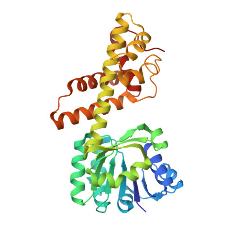
    3CKY

  • PubMed Abstract: 

    2-(Hydroxymethyl)glutarate dehydrogenase, the fourth enzyme of the anaerobic nicotinate fermentation pathway of Eubacterium barkeri, catalyzes the NADH-dependent conversion between (S)-2-formylglutarate and (S)-2-(hydroxymethyl)glutarate. As shown by its 2.3-A crystal structure, this enzyme is a novel member of the beta-hydroxyacid dehydrogenase family and adopts a tetrameric architecture with monomers interacting via their C-terminal catalytic domains. The NAD-binding domains protrude heterogeneously from the central, tetrameric core with domain rotation angles differing up to 12 degrees. Kinetic properties of the enzyme, including NADH inhibition constants, were determined. A strong NADH binding in contrast to weaker NAD(+) binding of the protein was inferred from fluorometrically determined binding constants for the dinucleotide cofactor. The data support either an Iso Ordered Bi Bi mechanism or a more common Ordered Bi Bi mechanism as found in other dehydrogenases.


  • Organizational Affiliation

    Philipps-Universität Marburg, Fachbereich Chemie, Hans-Meerwein-Strasse, D-35032 Marburg, Germany.


Macromolecules
Find similar proteins by:  (by identity cutoff)  |  3D Structure
Entity ID: 1
MoleculeChains Sequence LengthOrganismDetailsImage
2-hydroxymethyl glutarate dehydrogenase
A, B, C, D
301Eubacterium barkeriMutation(s): 0 
Gene Names: Hgd
EC: 1.1.1.291
UniProt
Find proteins for Q0QLF5 (Eubacterium barkeri)
Explore Q0QLF5 
Go to UniProtKB:  Q0QLF5
Entity Groups  
Sequence Clusters30% Identity50% Identity70% Identity90% Identity95% Identity100% Identity
UniProt GroupQ0QLF5
Sequence Annotations
Expand
  • Reference Sequence
Experimental Data & Validation

Experimental Data

  • Method: X-RAY DIFFRACTION
  • Resolution: 2.30 Å
  • R-Value Free: 0.222 
  • R-Value Work: 0.194 
  • R-Value Observed: 0.195 
  • Space Group: P 21 21 21
Unit Cell:
Length ( Å )Angle ( ˚ )
a = 82.978α = 90
b = 175.779β = 90
c = 83.709γ = 90
Software Package:
Software NamePurpose
SCALAdata scaling
PHASERphasing
REFMACrefinement
PDB_EXTRACTdata extraction
MOSFLMdata reduction

Structure Validation

View Full Validation Report


Entry History 

Deposition Data

Revision History  (Full details and data files)

  • Version 1.0: 2008-08-26
    Type: Initial release
  • Version 1.1: 2011-07-13
    Changes: Advisory, Version format compliance
  • Version 1.2: 2024-02-21
    Changes: Data collection, Database references, Refinement description