3CHX

Crystal structure of Methylosinus trichosporium OB3b particulate methane monooxygenase (pMMO)


Experimental Data Snapshot

  • Method: X-RAY DIFFRACTION
  • Resolution: 3.90 Å
  • R-Value Free: 0.377 
  • R-Value Work: 0.342 

wwPDB Validation   3D Report Full Report


This is version 1.2 of the entry. See complete history


Literature

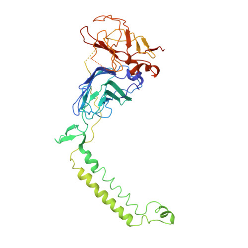
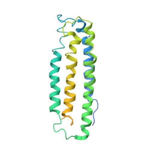
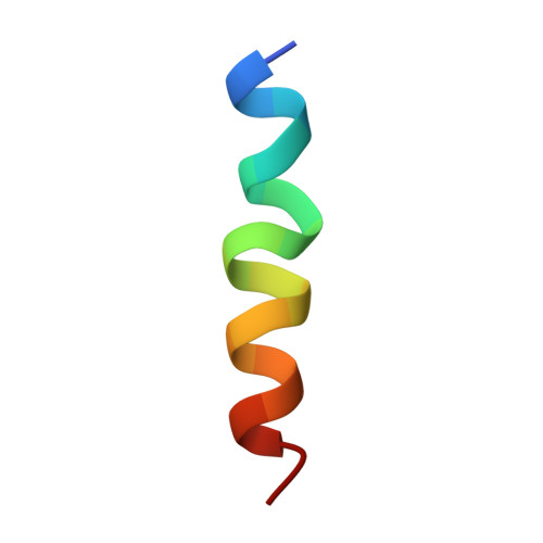
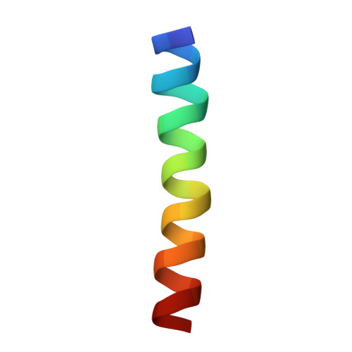
The metal centers of particulate methane monooxygenase from Methylosinus trichosporium OB3b.

Hakemian, A.S.Kondapalli, K.C.Telser, J.Hoffman, B.M.Stemmler, T.L.Rosenzweig, A.C.

(2008) Biochemistry 47: 6793-6801

  • DOI: https://doi.org/10.1021/bi800598h
  • Primary Citation of Related Structures:  
    3CHX

  • PubMed Abstract: 

    Particulate methane monooxygenase (pMMO) is a membrane-bound metalloenzyme that oxidizes methane to methanol in methanotrophic bacteria. The nature of the pMMO active site and the overall metal content are controversial, with spectroscopic and crystallographic data suggesting the presence of a mononuclear copper center, a dinuclear copper center, a trinuclear center, and a diiron center or combinations thereof. Most studies have focused on pMMO from Methylococcus capsulatus (Bath). pMMO from a second organism, Methylosinus trichosporium OB3b, has been purified and characterized by spectroscopic and crystallographic methods. Purified M. trichosporium OB3b pMMO contains approximately 2 copper ions per 100 kDa protomer. Electron paramagnetic resonance (EPR) spectroscopic parameters indicate that type 2 Cu(II) is present as two distinct species. Extended X-ray absorption fine structure (EXAFS) data are best fit with oxygen/nitrogen ligands and reveal a Cu-Cu interaction at 2.52 A. Correspondingly, X-ray crystallography of M. trichosporium OB3b pMMO shows a dinuclear copper center, similar to that observed previously in the crystal structure of M. capsulatus (Bath) pMMO. There are, however, significant differences between the pMMO structures from the two organisms. A mononuclear copper center present in M. capsulatus (Bath) pMMO is absent in M. trichosporium OB3b pMMO, whereas a metal center occupied by zinc in the M. capsulatus (Bath) pMMO structure is occupied by copper in M. trichosporium OB3b pMMO. These findings extend previous work on pMMO from M. capsulatus (Bath) and provide new insight into the functional importance of the different metal centers.


  • Organizational Affiliation

    Department of Biochemistry, Northwestern University, Evanston, Illinois 60208, USA.


Macromolecules
Find similar proteins by:  (by identity cutoff)  |  3D Structure
Entity ID: 1
MoleculeChains Sequence LengthOrganismDetailsImage
PmoBA,
F [auth E],
K [auth I]
392Methylosinus trichosporiumMutation(s): 0 
Gene Names: pmoB
Membrane Entity: Yes 
UniProt
Find proteins for Q9KX50 (Methylosinus trichosporium)
Explore Q9KX50 
Go to UniProtKB:  Q9KX50
Entity Groups  
Sequence Clusters30% Identity50% Identity70% Identity90% Identity95% Identity100% Identity
UniProt GroupQ9KX50
Sequence Annotations
Expand
  • Reference Sequence
Find similar proteins by:  (by identity cutoff)  |  3D Structure
Entity ID: 2
MoleculeChains Sequence LengthOrganismDetailsImage
PmoAB,
G [auth F],
L [auth J]
252Methylosinus trichosporiumMutation(s): 0 
Gene Names: pmoA
Membrane Entity: Yes 
UniProt
Find proteins for Q50541 (Methylosinus trichosporium)
Explore Q50541 
Go to UniProtKB:  Q50541
Entity Groups  
Sequence Clusters30% Identity50% Identity70% Identity90% Identity95% Identity100% Identity
UniProt GroupQ50541
Sequence Annotations
Expand
  • Reference Sequence
Find similar proteins by:  (by identity cutoff)  |  3D Structure
Entity ID: 3
MoleculeChains Sequence LengthOrganismDetailsImage
PmoCC,
H [auth G],
M [auth K]
256Methylosinus trichosporiumMutation(s): 0 
Gene Names: pmoC
Membrane Entity: Yes 
UniProt
Find proteins for Q9KX51 (Methylosinus trichosporium)
Explore Q9KX51 
Go to UniProtKB:  Q9KX51
Entity Groups  
Sequence Clusters30% Identity50% Identity70% Identity90% Identity95% Identity100% Identity
UniProt GroupQ9KX51
Sequence Annotations
Expand
  • Reference Sequence

Find similar proteins by:  Sequence   |   3D Structure  

Entity ID: 4
MoleculeChains Sequence LengthOrganismDetailsImage
20-residue peptideD,
I [auth H],
N [auth L]
20Methylosinus trichosporiumMutation(s): 0 
Sequence Annotations
Expand
  • Reference Sequence
Find similar proteins by:  (by identity cutoff)  |  3D Structure
Entity ID: 5
MoleculeChains Sequence LengthOrganismDetailsImage
26-residue peptideE [auth M],
J [auth N],
O
26Methylosinus trichosporiumMutation(s): 0 
Sequence Annotations
Expand
  • Reference Sequence
Experimental Data & Validation

Experimental Data

  • Method: X-RAY DIFFRACTION
  • Resolution: 3.90 Å
  • R-Value Free: 0.377 
  • R-Value Work: 0.342 
  • Space Group: P 21 21 21
Unit Cell:
Length ( Å )Angle ( ˚ )
a = 113.81α = 90
b = 184.07β = 90
c = 203.9γ = 90
Software Package:
Software NamePurpose
SCALAdata scaling
PHASERphasing
CNSrefinement
PDB_EXTRACTdata extraction
Blu-Icedata collection
XDSdata reduction

Structure Validation

View Full Validation Report


Entry History 

Deposition Data

Revision History  (Full details and data files)

  • Version 1.0: 2008-07-15
    Type: Initial release
  • Version 1.1: 2011-07-13
    Changes: Version format compliance
  • Version 1.2: 2023-08-30
    Changes: Data collection, Database references, Derived calculations, Refinement description