3CHH

Crystal Structure of Di-iron AurF


Experimental Data Snapshot

  • Method: X-RAY DIFFRACTION
  • Resolution: 2.00 Å
  • R-Value Free: 0.251 
  • R-Value Work: 0.200 
  • R-Value Observed: 0.203 

wwPDB Validation   3D Report Full Report


This is version 1.2 of the entry. See complete history


Literature

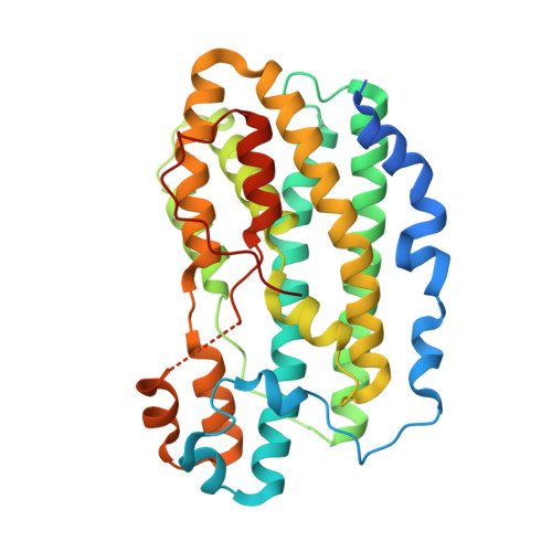
In vitro reconstitution and crystal structure of p-aminobenzoate N-oxygenase (AurF) involved in aureothin biosynthesis.

Choi, Y.S.Zhang, H.Brunzelle, J.S.Nair, S.K.Zhao, H.

(2008) Proc Natl Acad Sci U S A 105: 6858-6863

  • DOI: https://doi.org/10.1073/pnas.0712073105
  • Primary Citation of Related Structures:  
    3CHH, 3CHI, 3CHT, 3CHU

  • PubMed Abstract: 

    p-Aminobenzoate N-oxygenase (AurF) from Streptomyces thioluteus catalyzes the formation of unusual polyketide synthase starter unit p-nitrobenzoic acid (pNBA) from p-aminobenzoic acid (pABA) in the biosynthesis of antibiotic aureothin. AurF is a metalloenzyme, but its native enzymatic activity has not been demonstrated in vitro, and its catalytic mechanism is unclear. In addition, the nature of the cofactor remains a controversy. Here, we report the in vitro reconstitution of the AurF enzyme activity, the crystal structure of AurF in the oxidized state, and the cocrystal structure of AurF with its product pNBA. Our combined biochemical and structural analysis unequivocally indicates that AurF is a non-heme di-iron monooxygenase that catalyzes sequential oxidation of aminoarenes to nitroarenes via hydroxylamine and nitroso intermediates.


  • Organizational Affiliation

    Department of Chemical and Biomolecular Engineering, University of Illinois at Urbana-Champaign, 600 South Mathews Avenue, Urbana, IL 61801, USA.


Macromolecules
Find similar proteins by:  (by identity cutoff)  |  3D Structure
Entity ID: 1
MoleculeChains Sequence LengthOrganismDetailsImage
p-Aminobenzoate N-Oxygenase
A, B
336Streptomyces thioluteusMutation(s): 0 
Gene Names: aurF
UniProt
Find proteins for Q70KH9 (Streptomyces thioluteus)
Explore Q70KH9 
Go to UniProtKB:  Q70KH9
Entity Groups  
Sequence Clusters30% Identity50% Identity70% Identity90% Identity95% Identity100% Identity
UniProt GroupQ70KH9
Sequence Annotations
Expand
  • Reference Sequence
Experimental Data & Validation

Experimental Data

  • Method: X-RAY DIFFRACTION
  • Resolution: 2.00 Å
  • R-Value Free: 0.251 
  • R-Value Work: 0.200 
  • R-Value Observed: 0.203 
  • Space Group: P 21 21 21
Unit Cell:
Length ( Å )Angle ( ˚ )
a = 58.143α = 90
b = 77.188β = 90
c = 140.405γ = 90
Software Package:
Software NamePurpose
REFMACrefinement
MAR345dtbdata collection
HKL-2000data reduction
HKL-2000data scaling
SHARPphasing

Structure Validation

View Full Validation Report


Entry History 

Deposition Data

Revision History  (Full details and data files)

  • Version 1.0: 2008-05-27
    Type: Initial release
  • Version 1.1: 2011-07-13
    Changes: Advisory, Version format compliance
  • Version 1.2: 2024-02-21
    Changes: Data collection, Database references, Derived calculations