3CHG

The compatible solute-binding protein OpuAC from Bacillus subtilis in complex with DMSA


Experimental Data Snapshot

  • Method: X-RAY DIFFRACTION
  • Resolution: 2.80 Å
  • R-Value Free: 0.359 
  • R-Value Work: 0.285 
  • R-Value Observed: 0.289 

wwPDB Validation   3D Report Full Report


This is version 1.2 of the entry. See complete history


Literature

The Compatible-Solute-Binding Protein OpuAC from Bacillus subtilis: Ligand Binding, Site-Directed Mutagenesis, and Crystallographic Studies

Smits, S.H.J.Hoing, M.Lecher, J.Jebbar, M.Schmitt, L.Bremer, E.

(2008) J Bacteriol 190: 5663-5671

  • DOI: https://doi.org/10.1128/JB.00346-08
  • Primary Citation of Related Structures:  
    3CHG

  • PubMed Abstract: 

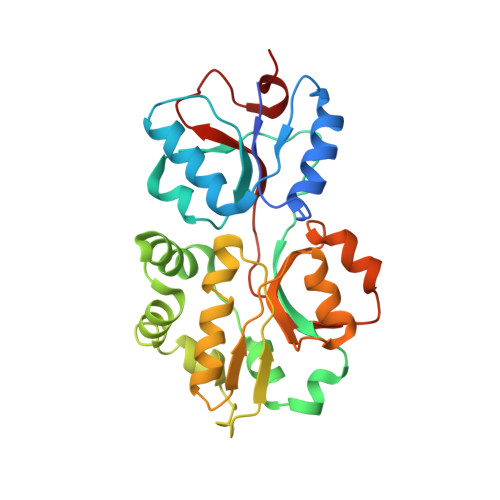
    In the soil bacterium Bacillus subtilis, five transport systems work in concert to mediate the import of various compatible solutes that counteract the deleterious effects of increases in the osmolarity of the environment. Among these five systems, the ABC transporter OpuA, which catalyzes the import of glycine betaine and proline betaine, has been studied in detail in the past. Here, we demonstrate that OpuA is capable of importing the sulfobetaine dimethylsulfonioacetate (DMSA). Since OpuA is a classic ABC importer that relies on a substrate-binding protein priming the transporter with specificity and selectivity, we analyzed the OpuA-binding protein OpuAC by structural and mutational means with respect to DMSA binding. The determined crystal structure of OpuAC in complex with DMSA at a 2.8-A resolution and a detailed mutational analysis of these residues revealed a hierarchy within the amino acids participating in substrate binding. This finding is different from those for other binding proteins that recognize compatible solutes. Furthermore, important principles that enable OpuAC to specifically bind various compatible solutes were uncovered.


  • Organizational Affiliation

    Institute of Biochemistry, Heinrich Heine University Düesseldorf, Universitätsstr. 1, 40225 Düsseldorf, Germany.


Macromolecules
Find similar proteins by:  (by identity cutoff)  |  3D Structure
Entity ID: 1
MoleculeChains Sequence LengthOrganismDetailsImage
Glycine betaine-binding proteinA [auth D],
B [auth A],
C [auth B],
D [auth C]
268Bacillus subtilisMutation(s): 0 
Gene Names: opuAC
UniProt
Find proteins for P46922 (Bacillus subtilis (strain 168))
Explore P46922 
Go to UniProtKB:  P46922
Entity Groups  
Sequence Clusters30% Identity50% Identity70% Identity90% Identity95% Identity100% Identity
UniProt GroupP46922
Sequence Annotations
Expand
  • Reference Sequence
Small Molecules
Experimental Data & Validation

Experimental Data

  • Method: X-RAY DIFFRACTION
  • Resolution: 2.80 Å
  • R-Value Free: 0.359 
  • R-Value Work: 0.285 
  • R-Value Observed: 0.289 
  • Space Group: P 1 21 1
Unit Cell:
Length ( Å )Angle ( ˚ )
a = 56.508α = 90
b = 150.606β = 104.54
c = 58.959γ = 90
Software Package:
Software NamePurpose
HKL-2000data collection
AMoREphasing
REFMACrefinement
XDSdata reduction
XSCALEdata scaling

Structure Validation

View Full Validation Report


Entry History 

Deposition Data

Revision History  (Full details and data files)

  • Version 1.0: 2008-08-12
    Type: Initial release
  • Version 1.1: 2011-07-13
    Changes: Version format compliance
  • Version 1.2: 2024-02-21
    Changes: Data collection, Database references, Derived calculations