3CH6

Crystal Structure of 11beta-HSD1 Double Mutant (L262R, F278E) Complexed with (3,3-dimethylpiperidin-1-yl)(6-(3-fluoro-4-methylphenyl)pyridin-2-yl)methanone


Experimental Data Snapshot

  • Method: X-RAY DIFFRACTION
  • Resolution: 2.35 Å
  • R-Value Free: 0.230 
  • R-Value Work: 0.187 
  • R-Value Observed: 0.188 

wwPDB Validation   3D Report Full Report


Ligand Structure Quality Assessment 


This is version 1.5 of the entry. See complete history


Literature

Pyridine amides as potent and selective inhibitors of 11beta-hydroxysteroid dehydrogenase type 1

Wang, H.Ruan, Z.Li, J.J.Simpkins, L.M.Smirk, R.A.Wu, S.C.Hutchins, R.D.Nirschl, D.S.Van Kirk, K.Cooper, C.B.Sutton, J.C.Ma, Z.Golla, R.Seethala, R.Salyan, M.E.Nayeem, A.Krystek, S.R.Sheriff, S.Camac, D.M.Morin, P.E.Carpenter, B.Robl, J.A.Zahler, R.Gordon, D.A.Hamann, L.G.

(2008) Bioorg Med Chem Lett 18: 3168-3172

  • DOI: https://doi.org/10.1016/j.bmcl.2008.04.069
  • Primary Citation of Related Structures:  
    3CH6

  • PubMed Abstract: 

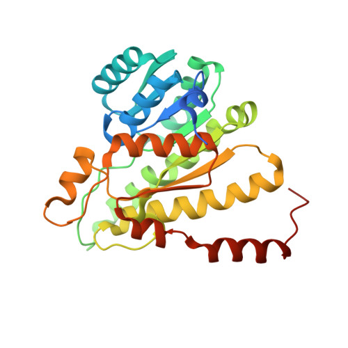
    Several series of pyridine amides were identified as selective and potent 11beta-HSD1 inhibitors. The most potent inhibitors feature 2,6- or 3,5-disubstitution on the pyridine core. Various linkers (CH(2)SO(2), CH(2)S, CH(2)O, S, O, N, bond) between the distal aryl and central pyridyl groups are tolerated, and lipophilic amide groups are generally favored. On the distal aryl group, a number of substitutions are well tolerated. A crystal structure was obtained for a complex between 11beta-HSD1 and the most potent inhibitor in this series.


  • Organizational Affiliation

    Bristol-Myers Squibb Research and Development, PO Box 5400, Princeton, NJ 08543-5400, USA. haixia.wang@bms.com


Macromolecules
Find similar proteins by:  (by identity cutoff)  |  3D Structure
Entity ID: 1
MoleculeChains Sequence LengthOrganismDetailsImage
Corticosteroid 11-beta-dehydrogenase isozyme 1A,
B,
C [auth D],
D [auth E]
286Homo sapiensMutation(s): 2 
Gene Names: HSD11B1HSD11HSD11L
EC: 1.1.1.146
UniProt & NIH Common Fund Data Resources
Find proteins for P28845 (Homo sapiens)
Explore P28845 
Go to UniProtKB:  P28845
PHAROS:  P28845
GTEx:  ENSG00000117594 
Entity Groups  
Sequence Clusters30% Identity50% Identity70% Identity90% Identity95% Identity100% Identity
UniProt GroupP28845
Sequence Annotations
Expand
  • Reference Sequence
Small Molecules
Binding Affinity Annotations 
IDSourceBinding Affinity
311 Binding MOAD:  3CH6 IC50: 0.1 (nM) from 1 assay(s)
BindingDB:  3CH6 IC50: 0.1 (nM) from 1 assay(s)
Experimental Data & Validation

Experimental Data

  • Method: X-RAY DIFFRACTION
  • Resolution: 2.35 Å
  • R-Value Free: 0.230 
  • R-Value Work: 0.187 
  • R-Value Observed: 0.188 
  • Space Group: P 21 21 21
Unit Cell:
Length ( Å )Angle ( ˚ )
a = 74.5α = 90
b = 94.3β = 90
c = 167γ = 90
Software Package:
Software NamePurpose
DENZOdata reduction
SCALEPACKdata scaling
TNTrefinement
PDB_EXTRACTdata extraction
HKL-2000data reduction
HKL-2000data scaling
AMoREphasing
BUSTER-TNTrefinement

Structure Validation

View Full Validation Report


Ligand Structure Quality Assessment 


Entry History 

Deposition Data

  • Released Date: 2008-06-10 
  • Deposition Author(s): Sheriff, S.

Revision History  (Full details and data files)

  • Version 1.0: 2008-06-10
    Type: Initial release
  • Version 1.1: 2011-07-13
    Changes: Version format compliance
  • Version 1.2: 2017-10-25
    Changes: Refinement description
  • Version 1.3: 2021-10-20
    Changes: Database references, Derived calculations
  • Version 1.4: 2024-02-21
    Changes: Data collection
  • Version 1.5: 2024-04-03
    Changes: Refinement description