3CFW

L-selectin lectin and EGF domains


Experimental Data Snapshot

  • Method: X-RAY DIFFRACTION
  • Resolution: 2.20 Å
  • R-Value Free: 0.248 
  • R-Value Work: 0.201 

wwPDB Validation   3D Report Full Report


Ligand Structure Quality Assessment 


This is version 2.1 of the entry. See complete history


Literature

Glycan Bound to the Selectin Low Affinity State Engages Glu-88 to Stabilize the High Affinity State under Force.

Mehta-D'souza, P.Klopocki, A.G.Oganesyan, V.Terzyan, S.Mather, T.Li, Z.Panicker, S.R.Zhu, C.McEver, R.P.

(2017) J Biol Chem 292: 2510-2518

  • DOI: https://doi.org/10.1074/jbc.M116.767186
  • Primary Citation of Related Structures:  
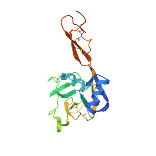
    3CFW

  • PubMed Abstract: 

    Selectin interactions with fucosylated glycan ligands mediate leukocyte rolling in the vasculature under shear forces. Crystal structures of P- and E-selectin suggest a two-state model in which ligand binding to the lectin domain closes loop 83-89 around the Ca 2+ coordination site, enabling Glu-88 to engage Ca 2+ and fucose. This triggers further allostery that opens the lectin/EGF domain hinge. The model posits that force accelerates transition from the bent (low affinity) to the extended (high affinity) state. However, transition intermediates have not been described, and the role of Glu-88 in force-assisted allostery has not been examined. Here we report the structure of the lectin and EGF domains of L-selectin bound to a fucose mimetic; that is, a terminal mannose on an N -glycan attached to a symmetry-related molecule. The structure is a transition intermediate where loop 83-89 closes to engage Ca 2+ and mannose without triggering allostery that opens the lectin/EGF domain hinge. We used three complementary assays to compare ligand binding to WT selectins and to E88D selectins that replaced Glu-88 with Asp. Soluble P-selectinE88D bound with an ∼9-fold lower affinity to PSGL-1, a physiological ligand, due to faster dissociation. Adhesion frequency experiments with a biomembrane force probe could not detect interactions of P-selectinE88D with PSGL-1. Cells expressing transmembrane P-selectinE88D or L-selectinE88D detached from immobilized ligands immediately after initiating flow. Cells expressing E-selectinE88D rolled but detached faster. Our data support a two-state model for selectins in which Glu-88 must engage ligand to trigger allostery that stabilizes the high affinity state under force.


  • Organizational Affiliation

    From the Cardiovascular Biology Research Program and.


Macromolecules
Find similar proteins by:  (by identity cutoff)  |  3D Structure
Entity ID: 1
MoleculeChains Sequence LengthOrganismDetailsImage
L-selectin164Homo sapiensMutation(s): 0 
Gene Names: L-selectinSELL
UniProt & NIH Common Fund Data Resources
Find proteins for P14151 (Homo sapiens)
Explore P14151 
Go to UniProtKB:  P14151
PHAROS:  P14151
GTEx:  ENSG00000188404 
Entity Groups  
Sequence Clusters30% Identity50% Identity70% Identity90% Identity95% Identity100% Identity
UniProt GroupP14151
Sequence Annotations
Expand
  • Reference Sequence
Oligosaccharides

Help

Entity ID: 2
MoleculeChains Length2D Diagram Glycosylation3D Interactions
alpha-D-mannopyranose-(1-3)-[beta-D-mannopyranose-(1-6)]alpha-D-mannopyranose-(1-4)-2-acetamido-2-deoxy-beta-D-glucopyranose-(1-4)-2-acetamido-2-deoxy-beta-D-glucopyranose
B
5N/A
Glycosylation Resources
GlyTouCan:  G08748CW
GlyCosmos:  G08748CW
GlyGen:  G08748CW
Small Molecules
Ligands 2 Unique
IDChains Name / Formula / InChI Key2D Diagram3D Interactions
NAG
Query on NAG

Download Ideal Coordinates CCD File 
C [auth A]2-acetamido-2-deoxy-beta-D-glucopyranose
C8 H15 N O6
OVRNDRQMDRJTHS-FMDGEEDCSA-N
CA
Query on CA

Download Ideal Coordinates CCD File 
D [auth A]CALCIUM ION
Ca
BHPQYMZQTOCNFJ-UHFFFAOYSA-N
Experimental Data & Validation

Experimental Data

  • Method: X-RAY DIFFRACTION
  • Resolution: 2.20 Å
  • R-Value Free: 0.248 
  • R-Value Work: 0.201 
  • Space Group: I 2 3
Unit Cell:
Length ( Å )Angle ( ˚ )
a = 118.477α = 90
b = 118.477β = 90
c = 118.477γ = 90
Software Package:
Software NamePurpose
HKL-2000data collection
AMoREphasing
CNSrefinement
DENZOdata reduction
SCALEPACKdata scaling

Structure Validation

View Full Validation Report


Ligand Structure Quality Assessment 


Entry History 

Deposition Data

Revision History  (Full details and data files)

  • Version 1.0: 2008-03-18
    Type: Initial release
  • Version 1.1: 2011-07-13
    Changes: Non-polymer description, Version format compliance
  • Version 1.2: 2017-02-22
    Changes: Database references
  • Version 2.0: 2020-07-29
    Type: Remediation
    Reason: Carbohydrate remediation
    Changes: Advisory, Atomic model, Data collection, Database references, Derived calculations, Structure summary
  • Version 2.1: 2023-08-30
    Changes: Advisory, Data collection, Database references, Refinement description, Structure summary