


Crystal structure of SC4828, a unique phosphatase from Streptomyces coelicolor.
Singer, A.U., Proudfoot, M., Brown, G., Xu, X., Chang, C., Zhang, H., Edwards, A.M., Joachimiak, A., Savchenko, A., Yakunin, A.F.To be published.
Experimental Data Snapshot
wwPDB Validation 3D Report Full Report

Entity ID: 1 | |||||
|---|---|---|---|---|---|
| Molecule | Chains | Sequence Length | Organism | Details | Image |
| Phosphatase SC4828 | 237 | Streptomyces coelicolor A3(2) | Mutation(s): 0 Gene Names: SCO5041, SCK7.14, SCK7.14c, gi:21223414 EC: 3.1.3.5 | 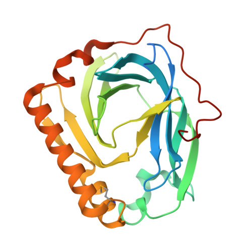 | |
UniProt | |||||
Find proteins for Q9FBN7 (Streptomyces coelicolor (strain ATCC BAA-471 / A3(2) / M145)) Explore Q9FBN7 Go to UniProtKB: Q9FBN7 | |||||
Entity Groups | |||||
| Sequence Clusters | 30% Identity50% Identity70% Identity90% Identity95% Identity100% Identity | ||||
| UniProt Group | Q9FBN7 | ||||
Sequence AnnotationsExpand | |||||
| |||||
| Ligands 2 Unique | |||||
|---|---|---|---|---|---|
| ID | Chains | Name / Formula / InChI Key | 2D Diagram | 3D Interactions | |
| MG Query on MG | B [auth A] | MAGNESIUM ION Mg JLVVSXFLKOJNIY-UHFFFAOYSA-N |  | ||
| NA Query on NA | C [auth A], D [auth A], E [auth A] | SODIUM ION Na FKNQFGJONOIPTF-UHFFFAOYSA-N |  | ||
| Modified Residues 1 Unique | |||||
|---|---|---|---|---|---|
| ID | Chains | Type | Formula | 2D Diagram | Parent |
| MSE Query on MSE | A | L-PEPTIDE LINKING | C5 H11 N O2 Se |  | MET |
| Length ( Å ) | Angle ( ˚ ) |
|---|---|
| a = 39.738 | α = 90 |
| b = 77.271 | β = 90 |
| c = 80.044 | γ = 90 |
| Software Name | Purpose |
|---|---|
| REFMAC | refinement |
| SBC-Collect | data collection |
| HKL-2000 | data reduction |
| HKL-2000 | data scaling |
| SHELXCD | phasing |
| SHELXE | model building |
RCSB PDB (citation) is hosted by
RCSB PDB is a member of the

