3CAK

X-ray structure of WT PTE with ethyl phosphate


Experimental Data Snapshot

  • Method: X-RAY DIFFRACTION
  • Resolution: 1.83 Å
  • R-Value Free: 0.196 
  • R-Value Work: 0.140 
  • R-Value Observed: 0.143 

wwPDB Validation   3D Report Full Report


Ligand Structure Quality Assessment 


This is version 1.4 of the entry. See complete history


Literature

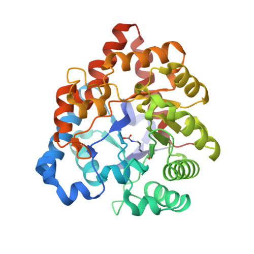
Structure of diethyl phosphate bound to the binuclear metal center of phosphotriesterase.

Kim, J.Tsai, P.C.Chen, S.L.Himo, F.Almo, S.C.Raushel, F.M.

(2008) Biochemistry 47: 9497-9504

  • DOI: https://doi.org/10.1021/bi800971v
  • Primary Citation of Related Structures:  
    2O4Q, 3CAK, 3CS2

  • PubMed Abstract: 

    The bacterial phosphotriesterase (PTE) from Pseudomonas diminuta catalyzes the hydrolysis of organophosphate esters at rates close to the diffusion limit. X-ray diffraction studies have shown that a binuclear metal center is positioned in the active site of PTE and that this complex is responsible for the activation of the nucleophilic water from solvent. In this paper, the three-dimensional structure of PTE was determined in the presence of the hydrolysis product, diethyl phosphate (DEP), and a product analogue, cacodylate. In the structure of the PTE-diethyl phosphate complex, the DEP product is found symmetrically bridging the two divalent cations. The DEP displaces the hydroxide from solvent that normally bridges the two divalent cations in structures determined in the presence or absence of substrate analogues. One of the phosphoryl oxygen atoms in the PTE-DEP complex is 2.0 A from the alpha-metal ion, while the other oxygen is 2.2 A from the beta-metal ion. The two metal ions are separated by a distance of 4.0 A. A similar structure is observed in the presence of cacodylate. Analogous complexes have previously been observed for the product complexes of isoaspartyl dipeptidase, d-aminoacylase, and dihydroorotase from the amidohydrolase superfamily of enzymes. The experimentally determined structure of the PTE-diethyl phosphate product complex is inconsistent with a recent proposal based upon quantum mechanical/molecular mechanical simulations which postulated the formation of an asymmetrical product complex bound exclusively to the beta-metal ion with a metal-metal separation of 5.3 A. This structure is also inconsistent with a chemical mechanism for substrate hydrolysis that utilizes the bridging hydroxide as a base to abstract a proton from a water molecule loosely associated with the alpha-metal ion. Density functional theory (DFT) calculations support a reaction mechanism that utilizes the bridging hydroxide as the direct nucleophile in the hydrolysis of organophosphate esters by PTE.


  • Organizational Affiliation

    Albert Einstein College of Medicine, 1300 Morris Park Avenue, Bronx, New York 10461, USA.


Macromolecules
Find similar proteins by:  (by identity cutoff)  |  3D Structure
Entity ID: 1
MoleculeChains Sequence LengthOrganismDetailsImage
Parathion hydrolase
A, B
331Brevundimonas diminutaMutation(s): 0 
Gene Names: opd
EC: 3.1.8.1
UniProt
Find proteins for P0A434 (Brevundimonas diminuta)
Explore P0A434 
Go to UniProtKB:  P0A434
Entity Groups  
Sequence Clusters30% Identity50% Identity70% Identity90% Identity95% Identity100% Identity
UniProt GroupP0A434
Sequence Annotations
Expand
  • Reference Sequence
Small Molecules
Ligands 4 Unique
IDChains Name / Formula / InChI Key2D Diagram3D Interactions
BTB
Query on BTB

Download Ideal Coordinates CCD File 
E [auth A],
I [auth B]
2-[BIS-(2-HYDROXY-ETHYL)-AMINO]-2-HYDROXYMETHYL-PROPANE-1,3-DIOL
C8 H19 N O5
OWMVSZAMULFTJU-UHFFFAOYSA-N
DPF
Query on DPF

Download Ideal Coordinates CCD File 
F [auth A]DIETHYL HYDROGEN PHOSPHATE
C4 H11 O4 P
UCQFCFPECQILOL-UHFFFAOYSA-N
EFS
Query on EFS

Download Ideal Coordinates CCD File 
J [auth B]ETHYL DIHYDROGEN PHOSPHATE
C2 H7 O4 P
ZJXZSIYSNXKHEA-UHFFFAOYSA-N
CO
Query on CO

Download Ideal Coordinates CCD File 
C [auth A],
D [auth A],
G [auth B],
H [auth B]
COBALT (II) ION
Co
XLJKHNWPARRRJB-UHFFFAOYSA-N
Modified Residues  1 Unique
IDChains TypeFormula2D DiagramParent
KCX
Query on KCX
A, B
L-PEPTIDE LINKINGC7 H14 N2 O4LYS
Experimental Data & Validation

Experimental Data

  • Method: X-RAY DIFFRACTION
  • Resolution: 1.83 Å
  • R-Value Free: 0.196 
  • R-Value Work: 0.140 
  • R-Value Observed: 0.143 
  • Space Group: P 1
Unit Cell:
Length ( Å )Angle ( ˚ )
a = 43.286α = 104.73
b = 45.338β = 93.4
c = 78.913γ = 97.65
Software Package:
Software NamePurpose
REFMACrefinement
HKL-2000data reduction
HKL-2000data scaling
MOLREPphasing

Structure Validation

View Full Validation Report


Ligand Structure Quality Assessment 


Entry History 

Deposition Data

Revision History  (Full details and data files)

  • Version 1.0: 2008-10-21
    Type: Initial release
  • Version 1.1: 2011-07-13
    Changes: Version format compliance
  • Version 1.2: 2017-10-25
    Changes: Refinement description
  • Version 1.3: 2023-08-30
    Changes: Data collection, Database references, Derived calculations, Refinement description
  • Version 1.4: 2023-11-15
    Changes: Data collection