3C6A

Crystal Structure of the RB49 gp17 nuclease domain


Experimental Data Snapshot

  • Method: X-RAY DIFFRACTION
  • Resolution: 1.16 Å
  • R-Value Free: 0.171 
  • R-Value Work: 0.134 
  • R-Value Observed: 0.134 

wwPDB Validation   3D Report Full Report


This is version 1.3 of the entry. See complete history


Literature

The structure of the phage T4 DNA packaging motor suggests a mechanism dependent on electrostatic forces.

Sun, S.Kondabagil, K.Draper, B.Alam, T.I.Bowman, V.D.Zhang, Z.Hegde, S.Fokine, A.Rossmann, M.G.Rao, V.B.

(2008) Cell 135: 1251-1262

  • DOI: https://doi.org/10.1016/j.cell.2008.11.015
  • Primary Citation of Related Structures:  
    3C6A, 3C6H, 3CPE, 3EZK

  • PubMed Abstract: 

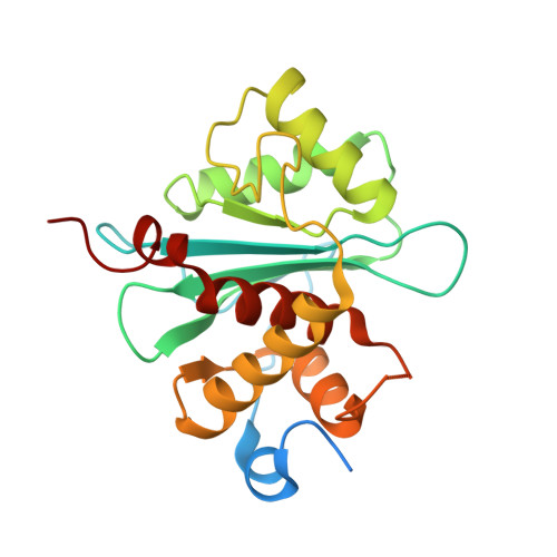
    Viral genomes are packaged into "procapsids" by powerful molecular motors. We report the crystal structure of the DNA packaging motor protein, gene product 17 (gp17), in bacteriophage T4. The structure consists of an N-terminal ATPase domain, which provides energy for compacting DNA, and a C-terminal nuclease domain, which terminates packaging. We show that another function of the C-terminal domain is to translocate the genome into the procapsid. The two domains are in close contact in the crystal structure, representing a "tensed state." A cryo-electron microscopy reconstruction of the T4 procapsid complexed with gp17 shows that the packaging motor is a pentamer and that the domains within each monomer are spatially separated, representing a "relaxed state." These structures suggest a mechanism, supported by mutational and other data, in which electrostatic forces drive the DNA packaging by alternating between tensed and relaxed states. Similar mechanisms may occur in other molecular motors.


  • Organizational Affiliation

    Department of Biological Sciences, Purdue University, 915 W. State Street, West Lafayette, IN 47907-2054, USA.


Macromolecules
Find similar proteins by:  (by identity cutoff)  |  3D Structure
Entity ID: 1
MoleculeChains Sequence LengthOrganismDetailsImage
Terminase large subunit232Escherichia phage RB49Mutation(s): 0 
Gene Names: 17
UniProt
Find proteins for Q9T1C3 (Escherichia phage RB49)
Explore Q9T1C3 
Go to UniProtKB:  Q9T1C3
Entity Groups  
Sequence Clusters30% Identity50% Identity70% Identity90% Identity95% Identity100% Identity
UniProt GroupQ9T1C3
Sequence Annotations
Expand
  • Reference Sequence
Small Molecules
Ligands 1 Unique
IDChains Name / Formula / InChI Key2D Diagram3D Interactions
MG
Query on MG

Download Ideal Coordinates CCD File 
B [auth A]MAGNESIUM ION
Mg
JLVVSXFLKOJNIY-UHFFFAOYSA-N
Experimental Data & Validation

Experimental Data

  • Method: X-RAY DIFFRACTION
  • Resolution: 1.16 Å
  • R-Value Free: 0.171 
  • R-Value Work: 0.134 
  • R-Value Observed: 0.134 
  • Space Group: P 21 21 2
Unit Cell:
Length ( Å )Angle ( ˚ )
a = 52.568α = 90
b = 125.163β = 90
c = 37.218γ = 90
Software Package:
Software NamePurpose
DENZOdata reduction
SCALEPACKdata scaling
SHARPphasing
SOLOMONphasing
SHELXrefinement
PDB_EXTRACTdata extraction
SHELXL-97refinement

Structure Validation

View Full Validation Report


Entry History 

Deposition Data

Revision History  (Full details and data files)

  • Version 1.0: 2009-01-13
    Type: Initial release
  • Version 1.1: 2011-07-13
    Changes: Version format compliance
  • Version 1.2: 2017-10-25
    Changes: Refinement description
  • Version 1.3: 2024-02-21
    Changes: Data collection, Database references, Derived calculations