3C3V

Crystal structure of peanut major allergen ara h 3


Experimental Data Snapshot

  • Method: X-RAY DIFFRACTION
  • Resolution: 1.73 Å
  • R-Value Free: 0.248 
  • R-Value Work: 0.186 
  • R-Value Observed: 0.189 

wwPDB Validation   3D Report Full Report


This is version 1.2 of the entry. See complete history


Literature

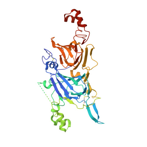
Crystal structure of Ara h 3, a major allergen in peanut.

Jin, T.Guo, F.Chen, Y.W.Howard, A.Zhang, Y.Z.

(2009) Mol Immunol 46: 1796-1804

  • DOI: https://doi.org/10.1016/j.molimm.2009.01.023
  • Primary Citation of Related Structures:  
    3C3V

  • PubMed Abstract: 

    The prevalence of food allergy has increased dramatically in recent years. Tremendous research progress has been made in understanding the pathophysiological mechanisms of allergy and in identifying and characterizing food allergens. Peanut is a major food allergen source and Ara h 3 is a major peanut allergen. Using overlapping short peptides, several linear IgE-binding epitopes in Ara h 3 have been defined before. However, the structure of Ara h 3 of the native allergen is not clear and information on conformational epitopes is lacking. Structural characterization of allergens is required for understanding the allergenicity of food allergens and for the development of immunotherapeutic agents. Previously, we have reported the crystallization of Ara h 3 purified from raw peanut. Here we report the crystal structure of Ara h 3 at 1.73A resolution. Mapping of the previously defined linear epitopes on the crystal structure of Ara h 3 indicated that linear epitopes with more solvent exposure were those indicated by the literature to react with more patient sera. The structure of Ara h 3 may be used to assess the importance of conformational epitopes in further investigations.


  • Organizational Affiliation

    Department of Biological, Chemical, and Physical Sciences, Illinois Institute of Technology, Chicago, IL 60616, United States.


Macromolecules
Find similar proteins by:  (by identity cutoff)  |  3D Structure
Entity ID: 1
MoleculeChains Sequence LengthOrganismDetailsImage
Arachin Arah3 isoform510Arachis hypogaeaMutation(s): 0 
UniProt
Find proteins for B5TYU1 (Arachis hypogaea)
Explore B5TYU1 
Go to UniProtKB:  B5TYU1
Entity Groups  
Sequence Clusters30% Identity50% Identity70% Identity90% Identity95% Identity100% Identity
UniProt GroupB5TYU1
Sequence Annotations
Expand
  • Reference Sequence
Small Molecules
Ligands 1 Unique
IDChains Name / Formula / InChI Key2D Diagram3D Interactions
NA
Query on NA

Download Ideal Coordinates CCD File 
B [auth A]SODIUM ION
Na
FKNQFGJONOIPTF-UHFFFAOYSA-N
Experimental Data & Validation

Experimental Data

  • Method: X-RAY DIFFRACTION
  • Resolution: 1.73 Å
  • R-Value Free: 0.248 
  • R-Value Work: 0.186 
  • R-Value Observed: 0.189 
  • Space Group: P 63 2 2
Unit Cell:
Length ( Å )Angle ( ˚ )
a = 144.929α = 90
b = 144.929β = 90
c = 86.924γ = 120
Software Package:
Software NamePurpose
DENZOdata reduction
SCALEPACKdata scaling
PHASERphasing
REFMACrefinement
PDB_EXTRACTdata extraction
HKL-2000data collection
HKL-2000data reduction
HKL-2000data scaling

Structure Validation

View Full Validation Report


Entry History 

Deposition Data

Revision History  (Full details and data files)

  • Version 1.0: 2009-02-24
    Type: Initial release
  • Version 1.1: 2011-07-13
    Changes: Version format compliance
  • Version 1.2: 2017-10-25
    Changes: Refinement description