3C3U

Crystal structure of AKR1C1 in complex with NADP and 3,5-dichlorosalicylic acid


Experimental Data Snapshot

  • Method: X-RAY DIFFRACTION
  • Resolution: 1.80 Å
  • R-Value Free: 0.218 
  • R-Value Work: 0.174 
  • R-Value Observed: 0.177 

wwPDB Validation   3D Report Full Report


Ligand Structure Quality Assessment 


This is version 1.2 of the entry. See complete history


Literature

Selectivity determinants of inhibitor binding to human 20alpha-hydroxysteroid dehydrogenase: crystal structure of the enzyme in ternary complex with coenzyme and the potent inhibitor 3,5-dichlorosalicylic acid

Dhagat, U.Endo, S.Sumii, R.Hara, A.El-Kabbani, O.

(2008) J Med Chem 51: 4844-4848

  • DOI: https://doi.org/10.1021/jm8003575
  • Primary Citation of Related Structures:  
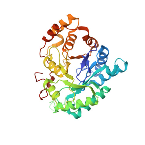
    3C3U

  • PubMed Abstract: 

    The crystal structure of human 20alpha-hydroxysteroid dehydrogenase (AKR1C1) in ternary complex with the coenzyme NADP (+) and the potent inhibitor 3,5-dichlorosalicylic acid was determined at a resolution of 1.8 A. The inhibitor is held in place by a network of hydrogen bonding interactions with the active site residues Tyr55, His117, and His222. The important role of the nonconserved residues Leu54, His222, Leu306, and Leu308 in inhibitor binding and selectivity was determined by site-directed mutagenesis.


  • Organizational Affiliation

    Medicinal Chemistry and Drug Action, Monash Institute of Pharmaceutical Sciences, Parkville, Victoria, Australia.


Macromolecules
Find similar proteins by:  (by identity cutoff)  |  3D Structure
Entity ID: 1
MoleculeChains Sequence LengthOrganismDetailsImage
Aldo-keto reductase family 1 member C1323Homo sapiensMutation(s): 0 
Gene Names: AKR1C1DDHDDH1
EC: 1.1.1.149
UniProt & NIH Common Fund Data Resources
Find proteins for Q04828 (Homo sapiens)
Explore Q04828 
Go to UniProtKB:  Q04828
PHAROS:  Q04828
GTEx:  ENSG00000187134 
Entity Groups  
Sequence Clusters30% Identity50% Identity70% Identity90% Identity95% Identity100% Identity
UniProt GroupQ04828
Sequence Annotations
Expand
  • Reference Sequence
Small Molecules
Ligands 3 Unique
IDChains Name / Formula / InChI Key2D Diagram3D Interactions
NAP
Query on NAP

Download Ideal Coordinates CCD File 
C [auth A]NADP NICOTINAMIDE-ADENINE-DINUCLEOTIDE PHOSPHATE
C21 H28 N7 O17 P3
XJLXINKUBYWONI-NNYOXOHSSA-N
C2U
Query on C2U

Download Ideal Coordinates CCD File 
D [auth A]3,5-dichloro-2-hydroxybenzoic acid
C7 H4 Cl2 O3
CNJGWCQEGROXEE-UHFFFAOYSA-N
ZN
Query on ZN

Download Ideal Coordinates CCD File 
B [auth A]ZINC ION
Zn
PTFCDOFLOPIGGS-UHFFFAOYSA-N
Binding Affinity Annotations 
IDSourceBinding Affinity
C2U BindingDB:  3C3U Ki: min: 5.9, max: 2800 (nM) from 8 assay(s)
IC50: 6 (nM) from 1 assay(s)
Binding MOAD:  3C3U Ki: 5.9 (nM) from 1 assay(s)
Experimental Data & Validation

Experimental Data

  • Method: X-RAY DIFFRACTION
  • Resolution: 1.80 Å
  • R-Value Free: 0.218 
  • R-Value Work: 0.174 
  • R-Value Observed: 0.177 
  • Space Group: P 1 21 1
Unit Cell:
Length ( Å )Angle ( ˚ )
a = 39.364α = 90
b = 84.236β = 91.26
c = 49.183γ = 90
Software Package:
Software NamePurpose
REFMACrefinement
HKL-2000data collection
HKL-2000data reduction
SCALEPACKdata scaling
MOLREPphasing

Structure Validation

View Full Validation Report


Ligand Structure Quality Assessment 


Entry History 

Deposition Data

Revision History  (Full details and data files)

  • Version 1.0: 2008-08-26
    Type: Initial release
  • Version 1.1: 2011-07-13
    Changes: Version format compliance
  • Version 1.2: 2023-11-01
    Changes: Data collection, Database references, Derived calculations, Refinement description