


The crystal structure of the tagatose-6-phosphate ketose/aldose isomerase from Escherichia coli.
Zhang, R., Skarina, T., Egorova, O., Savchenko, A., Edwards, A.M., Joachimiak, A.To be published.
Experimental Data Snapshot
wwPDB Validation 3D Report Full Report

Entity ID: 1 | |||||
|---|---|---|---|---|---|
| Molecule | Chains | Sequence Length | Organism | Details | Image |
| Putative tagatose-6-phosphate ketose/aldose isomerase | 384 | Escherichia coli K-12 | Mutation(s): 0 Gene Names: agaS, yraB, b3136, JW3105 | 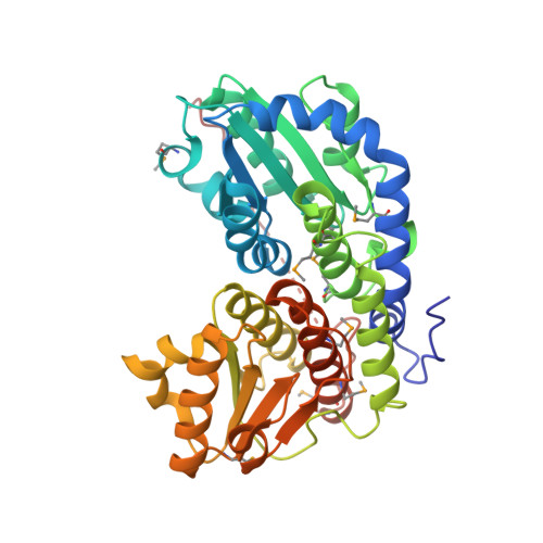 | |
UniProt | |||||
Find proteins for P42907 (Escherichia coli (strain K12)) Explore P42907 Go to UniProtKB: P42907 | |||||
Entity Groups | |||||
| Sequence Clusters | 30% Identity50% Identity70% Identity90% Identity95% Identity100% Identity | ||||
| UniProt Group | P42907 | ||||
Sequence AnnotationsExpand | |||||
| |||||
| Modified Residues 1 Unique | |||||
|---|---|---|---|---|---|
| ID | Chains | Type | Formula | 2D Diagram | Parent |
| MSE Query on MSE | A, B, C, D, E A, B, C, D, E, F | L-PEPTIDE LINKING | C5 H11 N O2 Se |  | MET |
| Length ( Å ) | Angle ( ˚ ) |
|---|---|
| a = 100.941 | α = 90 |
| b = 81.211 | β = 90.21 |
| c = 138.325 | γ = 90 |
| Software Name | Purpose |
|---|---|
| REFMAC | refinement |
| SBC-Collect | data collection |
| HKL-2000 | data reduction |
| HKL-2000 | data scaling |
| HKL-3000 | phasing |
RCSB PDB (citation) is hosted by
RCSB PDB is a member of the

