3BY8

Crystal Structure of the E.coli DcuS Sensor Domain


Experimental Data Snapshot

  • Method: X-RAY DIFFRACTION
  • Resolution: 1.45 Å
  • R-Value Free: 0.232 
  • R-Value Work: 0.219 

wwPDB Validation   3D Report Full Report


This is version 2.1 of the entry. See complete history


Literature

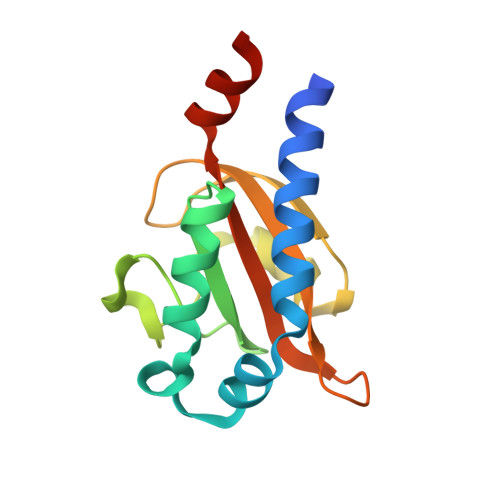
Crystal Structures of C4-Dicarboxylate Ligand Complexes with Sensor Domains of Histidine Kinases DcuS and DctB.

Cheung, J.Hendrickson, W.A.

(2008) J Biol Chem 283: 30256-30265

  • DOI: https://doi.org/10.1074/jbc.M805253200
  • Primary Citation of Related Structures:  
    3BY8, 3BY9

  • PubMed Abstract: 

    Two-component signaling systems allow bacteria to adapt to changing environments. Typically, a chemical or other stimulus is detected by the periplasmic sensor domain of a transmembrane histidine kinase sensor, which in turn relays a signal through a phosphotransfer cascade to the cognate cytoplasmic response regulator. Such systems lead ultimately to changes in gene expression or cell motility. Mechanisms of ligand binding and signal transduction through the cell membrane in histidine kinases are not fully understood. In an effort to further understand such processes, we have solved the crystal structures of the periplasmic sensor domains of Escherichia coli DcuS and of Vibrio cholerae DctB in complex with the respective cognate ligands, malate and succinate. Both proteins are involved in the regulation of the transport and metabolism of C(4)-dicarboxylates, but they are not highly related by sequence similarity. Our work reveals that despite disparate sizes, both structures contain a similar characteristic alpha/beta PDC (PhoQ-DcuS-CitA) sensor-domain fold and display similar modes of ligand binding, suggesting similar mechanisms of function.


  • Organizational Affiliation

    Department of Biochemistry and Molecular Biophysics, Columbia University, New York, New York 10032, USA.


Macromolecules
Find similar proteins by:  (by identity cutoff)  |  3D Structure
Entity ID: 1
MoleculeChains Sequence LengthOrganismDetailsImage
Sensor protein dcuS142Escherichia coliMutation(s): 0 
Gene Names: dcuSyjdH
EC: 2.7.13.3
UniProt
Find proteins for P0AEC8 (Escherichia coli (strain K12))
Explore P0AEC8 
Go to UniProtKB:  P0AEC8
Entity Groups  
Sequence Clusters30% Identity50% Identity70% Identity90% Identity95% Identity100% Identity
UniProt GroupP0AEC8
Sequence Annotations
Expand
  • Reference Sequence
Small Molecules
Ligands 1 Unique
IDChains Name / Formula / InChI Key2D Diagram3D Interactions
LMR
Query on LMR

Download Ideal Coordinates CCD File 
B [auth A](2S)-2-hydroxybutanedioic acid
C4 H6 O5
BJEPYKJPYRNKOW-REOHCLBHSA-N
Experimental Data & Validation

Experimental Data

  • Method: X-RAY DIFFRACTION
  • Resolution: 1.45 Å
  • R-Value Free: 0.232 
  • R-Value Work: 0.219 
  • Space Group: P 31 2 1
Unit Cell:
Length ( Å )Angle ( ˚ )
a = 86.111α = 90
b = 86.111β = 90
c = 35.207γ = 120
Software Package:
Software NamePurpose
DENZOdata reduction
SCALEPACKdata scaling
DMphasing
CNSrefinement
PDB_EXTRACTdata extraction

Structure Validation

View Full Validation Report


Entry History 

Deposition Data

Revision History  (Full details and data files)

  • Version 1.0: 2008-08-12
    Type: Initial release
  • Version 1.1: 2011-07-13
    Changes: Version format compliance
  • Version 1.2: 2017-10-25
    Changes: Refinement description
  • Version 2.0: 2021-04-21
    Changes: Atomic model, Data collection, Database references, Derived calculations, Non-polymer description, Structure summary
  • Version 2.1: 2024-02-21
    Changes: Data collection, Database references