3BY0

Crystal structure of Siderocalin (NGAL, Lipocalin 2) W79A-R81A complexed with Ferric Enterobactin


Experimental Data Snapshot

  • Method: X-RAY DIFFRACTION
  • Resolution: 2.57 Å
  • R-Value Free: 0.262 
  • R-Value Work: 0.248 
  • R-Value Observed: 0.249 

wwPDB Validation   3D Report Full Report


Ligand Structure Quality Assessment 


This is version 1.3 of the entry. See complete history


Literature

The siderocalin/enterobactin interaction: a link between mammalian immunity and bacterial iron transport.

Abergel, R.J.Clifton, M.C.Pizarro, J.C.Warner, J.A.Shuh, D.K.Strong, R.K.Raymond, K.N.

(2008) J Am Chem Soc 130: 11524-11534

  • DOI: https://doi.org/10.1021/ja803524w
  • Primary Citation of Related Structures:  
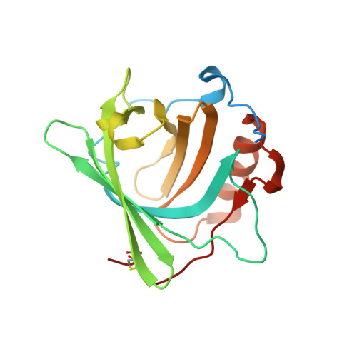
    3BY0, 3CBC

  • PubMed Abstract: 

    The siderophore enterobactin (Ent) is produced by enteric bacteria to mediate iron uptake. Ent scavenges iron and is taken up by the bacteria as the highly stable ferric complex [Fe (III)(Ent)] (3-). This complex is also a specific target of the mammalian innate immune system protein, Siderocalin (Scn), which acts as an antibacterial agent by specifically sequestering siderophores and their ferric complexes during infection. Recent literature suggesting that Scn may also be involved in cellular iron transport has increased the importance of understanding the mechanism of siderophore interception and clearance by Scn; Scn is observed to release iron in acidic endosomes and [Fe (III)(Ent)] (3-) is known to undergo a change from catecholate to salicylate coordination in acidic conditions, which is predicted to be sterically incompatible with the Scn binding pocket (also referred to as the calyx). To investigate the interactions between the ferric Ent complex and Scn at different pH values, two recombinant forms of Scn with mutations in three residues lining the calyx were prepared: Scn-W79A/R81A and Scn-Y106F. Binding studies and crystal structures of the Scn-W79A/R81A:[Fe (III)(Ent)] (3-) and Scn-Y106F:[Fe (III)(Ent)] (3-) complexes confirm that such mutations do not affect the overall conformation of the protein but do weaken significantly its affinity for [Fe (III)(Ent)] (3-). Fluorescence, UV-vis, and EXAFS spectroscopies were used to determine Scn/siderophore dissociation constants and to characterize the coordination mode of iron over a wide pH range, in the presence of both mutant proteins and synthetic salicylate analogues of Ent. While Scn binding hinders salicylate coordination transformation, strong acidification results in the release of iron and degraded siderophore. Iron release may therefore result from a combination of Ent degradation and coordination change.


  • Organizational Affiliation

    Department of Chemistry, University of California, Berkeley, California 94720-1460, USA.


Macromolecules
Find similar proteins by:  (by identity cutoff)  |  3D Structure
Entity ID: 1
MoleculeChains Sequence LengthOrganismDetailsImage
Neutrophil gelatinase-associated lipocalin
A, B, C
198Homo sapiensMutation(s): 3 
Gene Names: lcn2ngalhnl
UniProt & NIH Common Fund Data Resources
Find proteins for P80188 (Homo sapiens)
Explore P80188 
Go to UniProtKB:  P80188
PHAROS:  P80188
GTEx:  ENSG00000148346 
Entity Groups  
Sequence Clusters30% Identity50% Identity70% Identity90% Identity95% Identity100% Identity
UniProt GroupP80188
Sequence Annotations
Expand
  • Reference Sequence
Small Molecules
Ligands 5 Unique
IDChains Name / Formula / InChI Key2D Diagram3D Interactions
DBS
Query on DBS

Download Ideal Coordinates CCD File 
F [auth A],
N [auth C],
O [auth C]
2-(2,3-DIHYDROXY-BENZOYLAMINO)-3-HYDROXY-PROPIONIC ACID
C10 H11 N O6
VDTYHTVHFIIEIL-LURJTMIESA-N
DBH
Query on DBH

Download Ideal Coordinates CCD File 
G [auth A],
H [auth A],
K [auth B],
P [auth C]
2,3-DIHYDROXY-BENZOIC ACID
C7 H6 O4
GLDQAMYCGOIJDV-UHFFFAOYSA-N
SO4
Query on SO4

Download Ideal Coordinates CCD File 
E [auth A],
M [auth C]
SULFATE ION
O4 S
QAOWNCQODCNURD-UHFFFAOYSA-L
GOL
Query on GOL

Download Ideal Coordinates CCD File 
I [auth A],
Q [auth C],
R [auth C]
GLYCEROL
C3 H8 O3
PEDCQBHIVMGVHV-UHFFFAOYSA-N
FE
Query on FE

Download Ideal Coordinates CCD File 
D [auth A],
J [auth B],
L [auth C]
FE (III) ION
Fe
VTLYFUHAOXGGBS-UHFFFAOYSA-N
Experimental Data & Validation

Experimental Data

  • Method: X-RAY DIFFRACTION
  • Resolution: 2.57 Å
  • R-Value Free: 0.262 
  • R-Value Work: 0.248 
  • R-Value Observed: 0.249 
  • Space Group: P 41 21 2
Unit Cell:
Length ( Å )Angle ( ˚ )
a = 114.358α = 90
b = 114.358β = 90
c = 119.042γ = 90
Software Package:
Software NamePurpose
REFMACrefinement
HKL-2000data reduction
HKL-2000data scaling

Structure Validation

View Full Validation Report


Ligand Structure Quality Assessment 


Entry History 

Deposition Data

Revision History  (Full details and data files)

  • Version 1.0: 2008-09-09
    Type: Initial release
  • Version 1.1: 2011-07-13
    Changes: Non-polymer description, Version format compliance
  • Version 1.2: 2021-10-20
    Changes: Database references, Derived calculations
  • Version 1.3: 2023-08-30
    Changes: Data collection, Refinement description