3BXY

Crystal structure of tetrahydrodipicolinate N-succinyltransferase from E. coli


Experimental Data Snapshot

  • Method: X-RAY DIFFRACTION
  • Resolution: 2.00 Å
  • R-Value Free: 0.205 
  • R-Value Work: 0.175 
  • R-Value Observed: 0.176 

wwPDB Validation   3D Report Full Report


This is version 1.2 of the entry. See complete history


Literature

Structure of Escherichia coli tetrahydrodipicolinate N-succinyltransferase reveals the role of a conserved C-terminal helix in cooperative substrate binding.

Nguyen, L.Kozlov, G.Gehring, K.

(2008) FEBS Lett 582: 623-626

  • DOI: https://doi.org/10.1016/j.febslet.2008.01.032
  • Primary Citation of Related Structures:  
    3BXY

  • PubMed Abstract: 

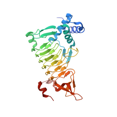
    Tetrahydrodipicolinate N-succinyltransferase is an enzyme present in many bacteria that catalyzes the first step of the succinylase pathway for the synthesis of meso-diaminopimelate and the amino acid L-lysine. Inhibition of the synthesis of meso-diaminopimelate, a component of peptidoglycan present in the cell wall of bacteria, is a potential route for the development of novel anti-bacterial agents. Here, we report the crystal structure of the DapD tetrahydrodipicolinate N-succinyltransferase from Escherichia coli at 2.0 A resolution. Comparison of the structure with the homologous enzyme from Mycobacterium bovis reveals the C-terminal helix undergoes a large rearrangement upon substrate binding, which contributes to cooperativity in substrate binding.


  • Organizational Affiliation

    Department of Biochemistry, McGill University, Montréal, 3655 Promenade Sir William Osler, Québec, Canada.


Macromolecules
Find similar proteins by:  (by identity cutoff)  |  3D Structure
Entity ID: 1
MoleculeChains Sequence LengthOrganismDetailsImage
2,3,4,5-tetrahydropyridine-2,6-dicarboxylate N-succinyltransferase285Escherichia coli O157:H7Mutation(s): 0 
Gene Names: dapD
EC: 2.3.1.117
UniProt
Find proteins for Q8X8Y7 (Escherichia coli O157:H7)
Explore Q8X8Y7 
Go to UniProtKB:  Q8X8Y7
Entity Groups  
Sequence Clusters30% Identity50% Identity70% Identity90% Identity95% Identity100% Identity
UniProt GroupQ8X8Y7
Sequence Annotations
Expand
  • Reference Sequence
Experimental Data & Validation

Experimental Data

  • Method: X-RAY DIFFRACTION
  • Resolution: 2.00 Å
  • R-Value Free: 0.205 
  • R-Value Work: 0.175 
  • R-Value Observed: 0.176 
  • Space Group: P 63
Unit Cell:
Length ( Å )Angle ( ˚ )
a = 102.924α = 90
b = 102.924β = 90
c = 69.229γ = 120
Software Package:
Software NamePurpose
REFMACrefinement
HKL-2000data collection
HKL-2000data reduction
HKL-2000data scaling
PHASERphasing

Structure Validation

View Full Validation Report


Entry History 

Deposition Data

Revision History  (Full details and data files)

  • Version 1.0: 2008-01-29
    Type: Initial release
  • Version 1.1: 2011-07-13
    Changes: Advisory, Refinement description, Source and taxonomy, Version format compliance
  • Version 1.2: 2023-08-30
    Changes: Data collection, Database references, Refinement description