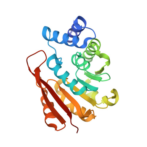
Crystal structures of human 108V and 108M catechol O-methyltransferase.
Rutherford, K., Le Trong, I., Stenkamp, R.E., Parson, W.W.(2008) J Mol Biol 380: 120-130
- PubMed: 18486144
- DOI: https://doi.org/10.1016/j.jmb.2008.04.040
- Primary Citation of Related Structures:
3BWM, 3BWY - PubMed Abstract:
Catechol O-methyltransferase (COMT) plays important roles in the metabolism of catecholamine neurotransmitters and catechol estrogens. The development of COMT inhibitors for use in the treatment of Parkinson's disease has been aided by crystallographic structures of the rat enzyme. However, the human and rat proteins have significantly different substrate specificities. Additionally, human COMT contains a common valine-methionine polymorphism at position 108. The methionine protein is less stable than the valine polymorph, resulting in decreased enzyme activity and protein levels in vivo. Here we describe the crystal structures of the 108V and 108M variants of the soluble form of human COMT bound with S-adenosylmethionine (SAM) and a substrate analog, 3,5-dinitrocatechol. The polymorphic residue 108 is located in the alpha5-beta3 loop, buried in a hydrophobic pocket approximately 16 A from the SAM-binding site. The 108V and 108M structures are very similar overall [RMSD of C(alpha) atoms between two structures (C(alpha) RMSD)=0.2 A], and the active-site residues are superposable, in accord with the observation that SAM stabilizes 108M COMT. However, the methionine side chain is packed more tightly within the polymorphic site and, consequently, interacts more closely with residues A22 (alpha2) and R78 (alpha4) than does valine. These interactions of the larger methionine result in a 0.7-A displacement in the backbone structure near residue 108, which propagates along alpha1 and alpha5 toward the SAM-binding site. Although the overall secondary structures of the human and rat proteins are very similar (C(alpha) RMSD=0.4 A), several nonconserved residues are present in the SAM-(I89M, I91M, C95Y) and catechol- (C173V, R201M, E202K) binding sites. The human protein also contains three additional solvent-exposed cysteine residues (C95, C173, C188) that may contribute to intermolecular disulfide bond formation and protein aggregation.
Organizational Affiliation:
Department of Biochemistry, University of Washington, Seattle, WA 98195, USA.