3BVO

Crystal structure of human co-chaperone protein HscB


Experimental Data Snapshot

  • Method: X-RAY DIFFRACTION
  • Resolution: 3.00 Å
  • R-Value Free: 0.288 
  • R-Value Work: 0.236 
  • R-Value Observed: 0.240 

wwPDB Validation   3D Report Full Report


This is version 1.2 of the entry. See complete history


Literature

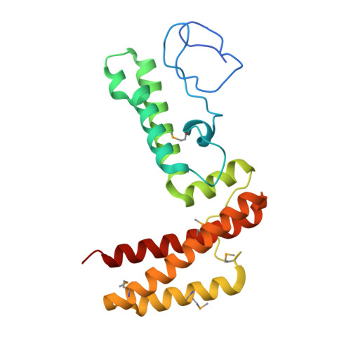
Structure of human J-type co-chaperone HscB reveals a tetracysteine metal-binding domain.

Bitto, E.Bingman, C.A.Bittova, L.Kondrashov, D.A.Bannen, R.M.Fox, B.G.Markley, J.L.Phillips, G.N.

(2008) J Biol Chem 283: 30184-30192

  • DOI: https://doi.org/10.1074/jbc.M804746200
  • Primary Citation of Related Structures:  
    3BVO

  • PubMed Abstract: 

    Iron-sulfur proteins play indispensable roles in a broad range of biochemical processes. The biogenesis of iron-sulfur proteins is a complex process that has become a subject of extensive research. The final step of iron-sulfur protein assembly involves transfer of an iron-sulfur cluster from a cluster-donor to a cluster-acceptor protein. This process is facilitated by a specialized chaperone system, which consists of a molecular chaperone from the Hsc70 family and a co-chaperone of the J-domain family. The 3.0 A crystal structure of a human mitochondrial J-type co-chaperone HscB revealed an L-shaped protein that resembles Escherichia coli HscB. The important difference between the two homologs is the presence of an auxiliary metal-binding domain at the N terminus of human HscB that coordinates a metal via the tetracysteine consensus motif CWXCX(9-13)FCXXCXXXQ. The domain is found in HscB homologs from animals and plants as well as in magnetotactic bacteria. The metal-binding site of the domain is structurally similar to that of rubredoxin and several zinc finger proteins containing rubredoxin-like knuckles. The normal mode analysis of HscB revealed that this L-shaped protein preferentially undergoes a scissors-like motion that correlates well with the conformational changes of human HscB observed in the crystals.


  • Organizational Affiliation

    Center for Eukaryotic Structural Genomics, University of Wisconsin-Madison, Madison, Wisconsin 53706-1544, USA.


Macromolecules
Find similar proteins by:  (by identity cutoff)  |  3D Structure
Entity ID: 1
MoleculeChains Sequence LengthOrganismDetailsImage
Co-chaperone protein HscB, mitochondrial precursor
A, B
207Homo sapiensMutation(s): 0 
Gene Names: HSCBDNAJC20HSC20
UniProt & NIH Common Fund Data Resources
Find proteins for Q8IWL3 (Homo sapiens)
Explore Q8IWL3 
Go to UniProtKB:  Q8IWL3
PHAROS:  Q8IWL3
GTEx:  ENSG00000100209 
Entity Groups  
Sequence Clusters30% Identity50% Identity70% Identity90% Identity95% Identity100% Identity
UniProt GroupQ8IWL3
Sequence Annotations
Expand
  • Reference Sequence
Small Molecules
Modified Residues  1 Unique
IDChains TypeFormula2D DiagramParent
MSE
Query on MSE
A, B
L-PEPTIDE LINKINGC5 H11 N O2 SeMET
Experimental Data & Validation

Experimental Data

  • Method: X-RAY DIFFRACTION
  • Resolution: 3.00 Å
  • R-Value Free: 0.288 
  • R-Value Work: 0.236 
  • R-Value Observed: 0.240 
  • Space Group: P 1 21 1
Unit Cell:
Length ( Å )Angle ( ˚ )
a = 63.644α = 90
b = 32.581β = 105.24
c = 114.362γ = 90
Software Package:
Software NamePurpose
DENZOdata reduction
SCALEPACKdata scaling
SHARPphasing
DMphasing
REFMACrefinement
PDB_EXTRACTdata extraction
MAR345data collection
HKL-2000data reduction
SHELXDphasing

Structure Validation

View Full Validation Report


Entry History 

Deposition Data

Revision History  (Full details and data files)

  • Version 1.0: 2008-01-15
    Type: Initial release
  • Version 1.1: 2011-07-13
    Changes: Advisory, Version format compliance
  • Version 1.2: 2017-10-25
    Changes: Refinement description