3BS6

1.8 Angstrom crystal structure of the periplasmic domain of the membrane insertase YidC


Experimental Data Snapshot

  • Method: X-RAY DIFFRACTION
  • Resolution: 1.80 Å
  • R-Value Free: 0.213 
  • R-Value Work: 0.178 
  • R-Value Observed: 0.179 

wwPDB Validation   3D Report Full Report


This is version 1.1 of the entry. See complete history


Literature

The Crystal Structure of the Periplasmic Domain of the Escherichia coli Membrane Protein Insertase YidC Contains a Substrate Binding Cleft

Ravaud, S.Stjepanovic, G.Wild, K.Sinning, I.

(2008) J Biol Chem 283: 9350-9358

  • DOI: https://doi.org/10.1074/jbc.M710493200
  • Primary Citation of Related Structures:  
    3BS6

  • PubMed Abstract: 

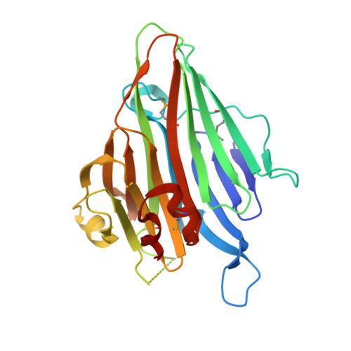
    In bacteria the biogenesis of inner membrane proteins requires targeting and insertion factors such as the signal recognition particle and the Sec translocon. YidC is an essential membrane protein involved in the insertion of inner membrane proteins together with the Sec translocon, but also as a separate entity. YidC of Escherichia coli is a member of the conserved YidC (in bacteria)/Oxa1 (in mitochondria)/Alb3 (in chloroplasts) protein family and contains six transmembrane segments and a large periplasmic domain (P1). We determined the crystal structure of the periplasmic domain of YidC from E. coli (P1D) at 1.8 A resolution. The structure of P1D shows the conserved beta-supersandwich fold of carbohydrate-binding proteins and an alpha-helical linker region at the C terminus that packs against the beta-supersandwich by a highly conserved interface. P1D exhibits an elongated cleft of similar architecture as found in the structural homologs. However, the electrostatic properties and molecular details of the cleft make it unlikely to interact with carbohydrate substrates. The cleft in P1D is occupied by a polyethylene glycol molecule suggesting an elongated peptide or acyl chain as a natural ligand. The region of P1D previously reported to interact with SecF maps to a surface area in the vicinity of the cleft. The conserved C-terminal region of the P1 domain was reported to be essential for the membrane insertase function of YidC. The analysis of this region suggests a role in membrane interaction and/or in the regulation of YidC interaction with binding partners.


  • Organizational Affiliation

    Biochemie-Zentrum der Universität Heidelberg, Im Neuenheimer Feld 328, Heidelberg, Germany.


Macromolecules
Find similar proteins by:  (by identity cutoff)  |  3D Structure
Entity ID: 1
MoleculeChains Sequence LengthOrganismDetailsImage
Inner membrane protein oxaA
A, B
280Escherichia coliMutation(s): 0 
Gene Names: oxaAyidC
UniProt
Find proteins for P25714 (Escherichia coli (strain K12))
Explore P25714 
Go to UniProtKB:  P25714
Entity Groups  
Sequence Clusters30% Identity50% Identity70% Identity90% Identity95% Identity100% Identity
UniProt GroupP25714
Sequence Annotations
Expand
  • Reference Sequence
Small Molecules
Ligands 5 Unique
IDChains Name / Formula / InChI Key2D Diagram3D Interactions
2PE
Query on 2PE

Download Ideal Coordinates CCD File 
N [auth B]NONAETHYLENE GLYCOL
C18 H38 O10
YZUUTMGDONTGTN-UHFFFAOYSA-N
PG4
Query on PG4

Download Ideal Coordinates CCD File 
H [auth A]TETRAETHYLENE GLYCOL
C8 H18 O5
UWHCKJMYHZGTIT-UHFFFAOYSA-N
PGE
Query on PGE

Download Ideal Coordinates CCD File 
I [auth A],
J [auth A],
O [auth B]
TRIETHYLENE GLYCOL
C6 H14 O4
ZIBGPFATKBEMQZ-UHFFFAOYSA-N
EDO
Query on EDO

Download Ideal Coordinates CCD File 
K [auth A]
L [auth A]
M [auth A]
P [auth B]
Q [auth B]
K [auth A],
L [auth A],
M [auth A],
P [auth B],
Q [auth B],
R [auth B],
S [auth B]
1,2-ETHANEDIOL
C2 H6 O2
LYCAIKOWRPUZTN-UHFFFAOYSA-N
CA
Query on CA

Download Ideal Coordinates CCD File 
C [auth A],
D [auth A],
E [auth A],
F [auth A],
G [auth A]
CALCIUM ION
Ca
BHPQYMZQTOCNFJ-UHFFFAOYSA-N
Modified Residues  1 Unique
IDChains TypeFormula2D DiagramParent
MSE
Query on MSE
A, B
L-PEPTIDE LINKINGC5 H11 N O2 SeMET
Experimental Data & Validation

Experimental Data

  • Method: X-RAY DIFFRACTION
  • Resolution: 1.80 Å
  • R-Value Free: 0.213 
  • R-Value Work: 0.178 
  • R-Value Observed: 0.179 
  • Space Group: C 1 2 1
Unit Cell:
Length ( Å )Angle ( ˚ )
a = 161.15α = 90
b = 55.64β = 101.12
c = 63.33γ = 90
Software Package:
Software NamePurpose
REFMACrefinement
PDB_EXTRACTdata extraction
ADSCdata collection
MOSFLMdata reduction
SCALAdata scaling
PHENIXphasing

Structure Validation

View Full Validation Report


Entry History 

Deposition Data

Revision History  (Full details and data files)

  • Version 1.0: 2008-02-12
    Type: Initial release
  • Version 1.1: 2011-07-13
    Changes: Version format compliance