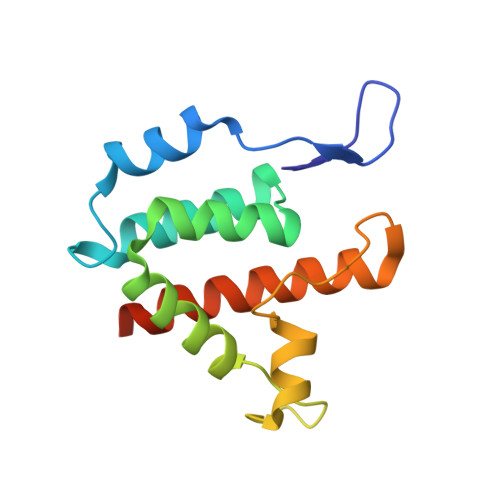
Structure of B-MLV capsid amino-terminal domain reveals key features of viral tropism, gag assembly and core formation
Mortuza, G.B., Dodding, M.P., Goldstone, D.C., Haire, L.F., Stoye, J.P., Taylor, I.A.(2008) J Mol Biol 376: 1493-1508
- PubMed: 18222469
- DOI: https://doi.org/10.1016/j.jmb.2007.12.043
- Primary Citation of Related Structures:
3BP9 - PubMed Abstract:
The Gag polyprotein is the major structural protein found in all classes of retroviruses. Interactions between Gag molecules control key events at several stages in the cycle of infection. In particular, the capsid (CA) domain of Gag mediates many of the protein-protein interactions that drive retrovirus assembly, maturation and disassembly. Moreover, in murine leukaemia virus (MLV), sequence variation in CA confers N and B tropism that determines susceptibility to the intracellular restriction factors Fv1n and Fv1b. We have determined the structure of the N-terminal domain (NtD) of CA from B-tropic MLV. A comparison of this structure with that of the NtD of CA from N-tropic MLV reveals that although the crystals belong to different space groups, CA monomers are packed with the same P6 hexagonal arrangement. Moreover, interhexamer crystal contacts between residues located at the periphery of the discs are conserved, indicating that switching of tropism does not result in large differences in the backbone conformation, nor does it alter the quaternary arrangement of the disc. We have also examined crystals of the N-tropic MLV CA containing both N- and C-terminal domains. In this case, the NtD hexamer is still present; however, the interhexamer spacing is increased and the conserved interhexamer contacts are absent. Investigation into the effects of mutation of residues that mediate interhexamer contacts reveals that amino acid substitutions at these positions cause severe defects in viral assembly, budding and Gag processing. Based on our crystal structures and mutational analysis, we propose that in MLV, interactions between the NtDs of CA are required for packing of Gag molecules in the early part of immature particle assembly. Moreover, we present a model where proteolytic cleavage at maturation results in migration of CA C-terminal domains into interstitial spaces between NtD hexamers. As a result, NtD-mediated interhexamer contacts present in the immature particle are displaced and the less densely packed lattice with increased hexamer-hexamer spacing characteristic of the viral core is produced.
Organizational Affiliation:
Division of Molecular Structure, National Institute for Medical Research, Ridgeway, Mill Hill, London NW7 1AA, UK.