3BI0

X-ray structure of human glutamate carboxypeptidase II (GCPII) in complex with a transition state analog of Glu-Glu


Experimental Data Snapshot

  • Method: X-RAY DIFFRACTION
  • Resolution: 1.67 Å
  • R-Value Free: 0.211 
  • R-Value Work: 0.183 
  • R-Value Observed: 0.184 

wwPDB Validation   3D Report Full Report


Ligand Structure Quality Assessment 


This is version 2.1 of the entry. See complete history


Literature

Structural basis of interactions between human glutamate carboxypeptidase II and its substrate analogs

Barinka, C.Hlouchova, K.Rovenska, M.Majer, P.Dauter, M.Hin, N.Ko, Y.S.Tsukamoto, T.Slusher, B.S.Konvalinka, J.Lubkowski, J.

(2008) J Mol Biol 376: 1438-1450

  • DOI: https://doi.org/10.1016/j.jmb.2007.12.066
  • Primary Citation of Related Structures:  
    3BHX, 3BI0, 3BI1

  • PubMed Abstract: 

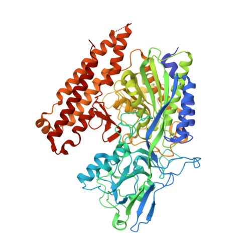
    Human glutamate carboxypeptidase II (GCPII) is involved in neuronal signal transduction and intestinal folate absorption by means of the hydrolysis of its two natural substrates, N-acetyl-aspartyl-glutamate and folyl-poly-gamma-glutamates, respectively. During the past years, tremendous efforts have been made toward the structural analysis of GCPII. Crystal structures of GCPII in complex with various ligands have provided insight into the binding of these ligands, particularly to the S1' site of the enzyme. In this article, we have extended structural characterization of GCPII to its S1 site by using dipeptide-based inhibitors that interact with both S1 and S1' sites of the enzyme. To this end, we have determined crystal structures of human GCPII in complex with phosphapeptide analogs of folyl-gamma-glutamate, aspartyl-glutamate, and gamma-glutamyl-glutamate, refined at 1.50, 1.60, and 1.67 A resolution, respectively. The S1 pocket of GCPII could be accurately defined and analyzed for the first time, and the data indicate the importance of Asn519, Arg463, Arg534, and Arg536 for recognition of the penultimate (i.e., P1) substrate residues. Direct interactions between the positively charged guanidinium groups of Arg534 and Arg536 and a P1 moiety of a substrate/inhibitor provide mechanistic explanation of GCPII preference for acidic dipeptides. Additionally, observed conformational flexibility of the Arg463 and Arg536 side chains likely regulates GCPII affinity toward different inhibitors and modulates GCPII substrate specificity. The biochemical experiments assessing the hydrolysis of several GCPII substrate derivatives modified at the P1 position, also included in this report, further complement and extend conclusions derived from the structural analysis. The data described here form an a solid foundation for the structurally aided design of novel low-molecular-weight GCPII inhibitors and imaging agents.


  • Organizational Affiliation

    Center for Cancer Research, National Cancer Institute, Frederick, MD 21702, USA. cyril@ncifcrf.gov


Macromolecules
Find similar proteins by:  (by identity cutoff)  |  3D Structure
Entity ID: 1
MoleculeChains Sequence LengthOrganismDetailsImage
Glutamate carboxypeptidase 2709Homo sapiensMutation(s): 0 
Gene Names: FOLH1FOLHNAALAD1PSMPSMA
EC: 3.4.17.21
UniProt & NIH Common Fund Data Resources
Find proteins for Q04609 (Homo sapiens)
Explore Q04609 
Go to UniProtKB:  Q04609
PHAROS:  Q04609
GTEx:  ENSG00000086205 
Entity Groups  
Sequence Clusters30% Identity50% Identity70% Identity90% Identity95% Identity100% Identity
UniProt GroupQ04609
Sequence Annotations
Expand
  • Reference Sequence
Oligosaccharides

Help

Entity ID: 2
MoleculeChains Length2D Diagram Glycosylation3D Interactions
2-acetamido-2-deoxy-beta-D-glucopyranose-(1-4)-2-acetamido-2-deoxy-beta-D-glucopyranose
B, C, D
2N-Glycosylation
Glycosylation Resources
GlyTouCan:  G42666HT
GlyCosmos:  G42666HT
GlyGen:  G42666HT
Entity ID: 3
MoleculeChains Length2D Diagram Glycosylation3D Interactions
alpha-D-mannopyranose-(1-3)-beta-D-mannopyranose-(1-4)-2-acetamido-2-deoxy-beta-D-glucopyranose-(1-4)-2-acetamido-2-deoxy-beta-D-glucopyranose
E
4N-Glycosylation
Glycosylation Resources
GlyTouCan:  G81315DD
GlyCosmos:  G81315DD
GlyGen:  G81315DD
Small Molecules
Ligands 5 Unique
IDChains Name / Formula / InChI Key2D Diagram3D Interactions
BIX
Query on BIX

Download Ideal Coordinates CCD File 
M [auth A](2S)-2-{[(S)-[(3S)-3-amino-3-carboxypropyl](hydroxy)phosphoryl]methyl}pentanedioic acid
C10 H18 N O8 P
YLHAQDFYEKJARV-RQJHMYQMSA-N
NAG
Query on NAG

Download Ideal Coordinates CCD File 
F [auth A],
G [auth A],
H [auth A]
2-acetamido-2-deoxy-beta-D-glucopyranose
C8 H15 N O6
OVRNDRQMDRJTHS-FMDGEEDCSA-N
ZN
Query on ZN

Download Ideal Coordinates CCD File 
I [auth A],
J [auth A]
ZINC ION
Zn
PTFCDOFLOPIGGS-UHFFFAOYSA-N
CA
Query on CA

Download Ideal Coordinates CCD File 
K [auth A]CALCIUM ION
Ca
BHPQYMZQTOCNFJ-UHFFFAOYSA-N
CL
Query on CL

Download Ideal Coordinates CCD File 
L [auth A]CHLORIDE ION
Cl
VEXZGXHMUGYJMC-UHFFFAOYSA-M
Binding Affinity Annotations 
IDSourceBinding Affinity
BIX Binding MOAD:  3BI0 IC50: 14 (nM) from 1 assay(s)
EPE PDBBind:  3BI0 IC50: 14 (nM) from 1 assay(s)
Experimental Data & Validation

Experimental Data

  • Method: X-RAY DIFFRACTION
  • Resolution: 1.67 Å
  • R-Value Free: 0.211 
  • R-Value Work: 0.183 
  • R-Value Observed: 0.184 
  • Space Group: I 2 2 2
Unit Cell:
Length ( Å )Angle ( ˚ )
a = 102.405α = 90
b = 130.797β = 90
c = 159.14γ = 90
Software Package:
Software NamePurpose
REFMACrefinement
HKL-2000data collection
HKL-2000data reduction
HKL-2000data scaling

Structure Validation

View Full Validation Report


Ligand Structure Quality Assessment 


Entry History 

Deposition Data

Revision History  (Full details and data files)

  • Version 1.0: 2008-01-08
    Type: Initial release
  • Version 1.1: 2011-07-13
    Changes: Non-polymer description, Version format compliance
  • Version 2.0: 2020-07-29
    Type: Remediation
    Reason: Carbohydrate remediation
    Changes: Atomic model, Data collection, Database references, Derived calculations, Structure summary
  • Version 2.1: 2023-08-30
    Changes: Data collection, Database references, Refinement description, Structure summary