3BFJ

Crystal structure analysis of 1,3-propanediol oxidoreductase


Experimental Data Snapshot

  • Method: X-RAY DIFFRACTION
  • Resolution: 2.70 Å
  • R-Value Free: 0.251 
  • R-Value Work: 0.203 
  • R-Value Observed: 0.205 

wwPDB Validation   3D Report Full Report


This is version 1.3 of the entry. See complete history


Literature

1,3-propanediol dehydrogenase from Klebsiella pneumoniae: decameric quaternary structure and possible subunit cooperativity

Marcal, D.Rego, A.T.Carrondo, M.A.Enguita, F.J.

(2009) J Bacteriol 191: 1143-1151

  • DOI: https://doi.org/10.1128/JB.01077-08
  • Primary Citation of Related Structures:  
    3BFJ

  • PubMed Abstract: 

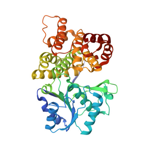
    Klebsiella pneumoniae is a nosocomial pathogen frequently isolated from opportunistic infections, especially in clinical environments. In spite of its potential pathogenicity, this microorganism has several metabolic potentials that could be used in biotechnology applications. K. pneumoniae is able to metabolize glycerol as a sole source of carbon and energy. 1,3-Propanediol dehydrogenase is the core of the metabolic pathway for the use of glycerol. We have determined the crystallographic structure of 1,3-propanediol dehydrogenase, a type III Fe-NAD-dependent alcohol dehydrogenase, at 2.7-A resolution. The structure of the enzyme monomer is closely related to that of other alcohol dehydrogenases. The overall arrangement of the enzyme showed a decameric structure, formed by a pentamer of dimers, which is the catalytic form of the enzyme. Dimers are associated by strong ionic interactions that are responsible for the highly stable in vivo packing of the enzyme. Kinetic properties of the enzyme as determined in the article would suggest that this decameric arrangement is related to the cooperativity between monomers.


  • Organizational Affiliation

    Instituto de Tecnologia Química e Biológica, Universidade Nova de Lisboa, 2781-901 Oeiras, Portugal.


Macromolecules
Find similar proteins by:  (by identity cutoff)  |  3D Structure
Entity ID: 1
MoleculeChains Sequence LengthOrganismDetailsImage
1,3-propanediol oxidoreductase
A, B, C, D, E
A, B, C, D, E, F, G, H, I, J, K, L, M, N, O, P, Q, R, S, T
387Klebsiella pneumoniaeMutation(s): 0 
Gene Names: dhaT
EC: 1.1.1.202
UniProt
Find proteins for Q7WRJ3 (Klebsiella pneumoniae)
Explore Q7WRJ3 
Go to UniProtKB:  Q7WRJ3
Entity Groups  
Sequence Clusters30% Identity50% Identity70% Identity90% Identity95% Identity100% Identity
UniProt GroupQ7WRJ3
Sequence Annotations
Expand
  • Reference Sequence
Small Molecules
Ligands 1 Unique
IDChains Name / Formula / InChI Key2D Diagram3D Interactions
FE2
Query on FE2

Download Ideal Coordinates CCD File 
AA [auth G]
BA [auth H]
CA [auth I]
DA [auth J]
EA [auth K]
AA [auth G],
BA [auth H],
CA [auth I],
DA [auth J],
EA [auth K],
FA [auth L],
GA [auth M],
HA [auth N],
IA [auth O],
JA [auth P],
KA [auth Q],
LA [auth R],
MA [auth S],
NA [auth T],
U [auth A],
V [auth B],
W [auth C],
X [auth D],
Y [auth E],
Z [auth F]
FE (II) ION
Fe
CWYNVVGOOAEACU-UHFFFAOYSA-N
Experimental Data & Validation

Experimental Data

  • Method: X-RAY DIFFRACTION
  • Resolution: 2.70 Å
  • R-Value Free: 0.251 
  • R-Value Work: 0.203 
  • R-Value Observed: 0.205 
  • Space Group: P 1 21 1
Unit Cell:
Length ( Å )Angle ( ˚ )
a = 91.937α = 90
b = 226.611β = 92.91
c = 232.627γ = 90
Software Package:
Software NamePurpose
REFMACrefinement
MOSFLMdata reduction
SCALAdata scaling
PHASERphasing

Structure Validation

View Full Validation Report


Entry History 

Deposition Data

Revision History  (Full details and data files)

  • Version 1.0: 2008-11-25
    Type: Initial release
  • Version 1.1: 2011-07-13
    Changes: Advisory, Refinement description, Version format compliance
  • Version 1.2: 2018-01-24
    Changes: Structure summary
  • Version 1.3: 2024-02-21
    Changes: Data collection, Database references, Derived calculations, Refinement description