3BF6

Thrombin:suramin complex


Experimental Data Snapshot

  • Method: X-RAY DIFFRACTION
  • Resolution: 2.50 Å
  • R-Value Free: 0.282 
  • R-Value Work: 0.228 
  • R-Value Observed: 0.231 

wwPDB Validation   3D Report Full Report


Ligand Structure Quality Assessment 


This is version 1.2 of the entry. See complete history


Literature

Structural and thermodynamic analysis of thrombin:suramin interaction in solution and crystal phases.

Lima, L.M.Becker, C.F.Giesel, G.M.Marques, A.F.Cargnelutti, M.T.de Oliveira Neto, M.Queiroz Monteiro, R.Verli, H.Polikarpov, I.

(2009) Biochim Biophys Acta 1794: 873-881

  • DOI: https://doi.org/10.1016/j.bbapap.2009.03.011
  • Primary Citation of Related Structures:  
    2H9T, 3BF6

  • PubMed Abstract: 

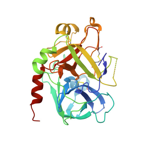
    Suramin is a hexasulfonated naphthylurea which has been recently characterized as a non-competitive inhibitor of human alpha-thrombin activity over fibrinogen, although its binding site and mode of interaction with the enzyme remain elusive. Here, we determined two X-ray structure of the thrombin:suramin complex, refined at 2.4 A resolution. While a single thrombin:suramin complex was found in the asymmetric unit cell of the crystal, some of the crystallographic contacts with symmetrically related molecules are mediated by both the enzyme and the ligand. Molecular dynamics simulations with the 1:1 complex demonstrate a large rearrangement of suramin in the complex, but with the protein scaffold and the more extensive protein-ligand regions keep unchanged. Small-angle X-ray scattering measurements at high micromolar concentration demonstrate a suramin-induced dimerization of the enzyme. These data indicating a dissimilar binding mode in the monomeric and oligomeric states, with a monomeric, 1:1 complex to be more likely to exist at the thrombin physiological, nanomolar concentration range. Collectively, close understanding on the structural basis for interaction is given which might establish a basis for design of suramin analogues targeting thrombin.


  • Organizational Affiliation

    Faculdade de Farmácia, Universidade Federal do Rio de Janeiro, 21941-590, Rio de Janeiro, RJ, Brazil. mauricio@pharma.ufrj.br


Macromolecules
Find similar proteins by:  (by identity cutoff)  |  3D Structure
Entity ID: 1
MoleculeChains Sequence LengthOrganismDetailsImage
Thrombin, LIGHT CHAINA [auth L]36Homo sapiensMutation(s): 0 
EC: 3.4.21.5
UniProt & NIH Common Fund Data Resources
Find proteins for P00734 (Homo sapiens)
Explore P00734 
Go to UniProtKB:  P00734
PHAROS:  P00734
GTEx:  ENSG00000180210 
Entity Groups  
Sequence Clusters30% Identity50% Identity70% Identity90% Identity95% Identity100% Identity
UniProt GroupP00734
Sequence Annotations
Expand
  • Reference Sequence
Find similar proteins by:  (by identity cutoff)  |  3D Structure
Entity ID: 2
MoleculeChains Sequence LengthOrganismDetailsImage
Thrombin, HEAVY CHAINB [auth H]259Homo sapiensMutation(s): 0 
EC: 3.4.21.5
UniProt & NIH Common Fund Data Resources
Find proteins for P00734 (Homo sapiens)
Explore P00734 
Go to UniProtKB:  P00734
PHAROS:  P00734
GTEx:  ENSG00000180210 
Entity Groups  
Sequence Clusters30% Identity50% Identity70% Identity90% Identity95% Identity100% Identity
UniProt GroupP00734
Sequence Annotations
Expand
  • Reference Sequence

Find similar proteins by:  Sequence   |   3D Structure  

Entity ID: 3
MoleculeChains Sequence LengthOrganismDetailsImage
PHE-PRO-ARGC [auth I]3N/AMutation(s): 0 
Sequence Annotations
Expand
  • Reference Sequence
Small Molecules
Ligands 1 Unique
IDChains Name / Formula / InChI Key2D Diagram3D Interactions
SVR
Query on SVR

Download Ideal Coordinates CCD File 
D [auth H]8,8'-[CARBONYLBIS[IMINO-3,1-PHENYLENECARBONYLIMINO(4-METHYL-3,1-PHENYLENE)CARBONYLIMINO]]BIS-1,3,5-NAPHTHALENETRISULFON IC ACID
C51 H40 N6 O23 S6
FIAFUQMPZJWCLV-UHFFFAOYSA-N
Binding Affinity Annotations 
IDSourceBinding Affinity
SVR Binding MOAD:  3BF6 IC50: 4.00e+4 (nM) from 1 assay(s)
Experimental Data & Validation

Experimental Data

  • Method: X-RAY DIFFRACTION
  • Resolution: 2.50 Å
  • R-Value Free: 0.282 
  • R-Value Work: 0.228 
  • R-Value Observed: 0.231 
  • Space Group: P 21 21 21
Unit Cell:
Length ( Å )Angle ( ˚ )
a = 44.381α = 90
b = 73.471β = 90
c = 114.707γ = 90
Software Package:
Software NamePurpose
MOSFLMdata reduction
SCALAdata scaling
REFMACrefinement
PDB_EXTRACTdata extraction
MAR345dtbdata collection
REFMACphasing

Structure Validation

View Full Validation Report


Ligand Structure Quality Assessment 


Entry History 

Deposition Data

Revision History  (Full details and data files)

  • Version 1.0: 2007-12-25
    Type: Initial release
  • Version 1.1: 2011-07-13
    Changes: Version format compliance
  • Version 1.2: 2015-07-15
    Changes: Structure summary