3BEN

Structure of N-(12-imidazolyl-dodecanoyl)-L-leucine inhibitor bound to the heme domain of Cytochrome P450-BM3


Experimental Data Snapshot

  • Method: X-RAY DIFFRACTION
  • Resolution: 1.65 Å
  • R-Value Free: 0.191 
  • R-Value Work: 0.161 
  • R-Value Observed: 0.162 

wwPDB Validation   3D Report Full Report


Ligand Structure Quality Assessment 


This is version 1.2 of the entry. See complete history


Literature

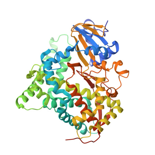
Crystal structure of inhibitor-bound P450BM-3 reveals open conformation of substrate access channel.

Haines, D.C.Chen, B.Tomchick, D.R.Bondlela, M.Hegde, A.Machius, M.Peterson, J.A.

(2008) Biochemistry 47: 3662-3670

  • DOI: https://doi.org/10.1021/bi7023964
  • Primary Citation of Related Structures:  
    3BEN

  • PubMed Abstract: 

    P450BM-3 is an extensively studied P450 cytochrome that is naturally fused to a cytochrome P450 reductase domain. Crystal structures of the heme domain of this enzyme have previously generated many insights into features of P450 structure, substrate binding specificity, and conformational changes that occur on substrate binding. Although many P450s are inhibited by imidazole, this compound does not effectively inhibit P450BM-3. Omega-imidazolyl fatty acids have previously been found to be weak inhibitors of the enzyme and show some unusual cooperativity with the substrate lauric acid. We set out to improve the properties of these inhibitors by attaching the omega-imidazolyl fatty acid to the nitrogen of an amino acid group, a tactic that we used previously to increase the potency of substrates. The resulting inhibitors were significantly more potent than their parent compounds lacking the amino acid group. A crystal structure of one of the new inhibitors bound to the heme domain of P450BM-3 reveals that the mode of interaction of the amino acid group with the enzyme is different from that previously observed for acyl amino acid substrates. Further, required movements of residues in the active site to accommodate the imidazole group provide an explanation for the low affinity of imidazole itself. Finally, the previously observed cooperativity with lauric acid is explained by a surprisingly open substrate-access channel lined with hydrophobic residues that could potentially accommodate lauric acid in addition to the inhibitor itself.


  • Organizational Affiliation

    Department of Chemistry, The University of Texas at Dallas, Dallas, Texas 75083-0688, USA.


Macromolecules
Find similar proteins by:  (by identity cutoff)  |  3D Structure
Entity ID: 1
MoleculeChains Sequence LengthOrganismDetailsImage
Cytochrome P450 102
A, B
470N/AMutation(s): 0 
Gene Names: CYP102A1cyp102cyp102A1
EC: 1.14.14.1
UniProt
Find proteins for P14779 (Priestia megaterium (strain ATCC 14581 / DSM 32 / CCUG 1817 / JCM 2506 / NBRC 15308 / NCIMB 9376 / NCTC 10342 / NRRL B-14308 / VKM B-512 / Ford 19))
Explore P14779 
Go to UniProtKB:  P14779
Entity Groups  
Sequence Clusters30% Identity50% Identity70% Identity90% Identity95% Identity100% Identity
UniProt GroupP14779
Sequence Annotations
Expand
  • Reference Sequence
Small Molecules
Ligands 4 Unique
IDChains Name / Formula / InChI Key2D Diagram3D Interactions
HEM
Query on HEM

Download Ideal Coordinates CCD File 
C [auth A],
E [auth B]
PROTOPORPHYRIN IX CONTAINING FE
C34 H32 Fe N4 O4
KABFMIBPWCXCRK-RGGAHWMASA-L
LEH
Query on LEH

Download Ideal Coordinates CCD File 
D [auth A],
F [auth B]
N-[12-(1H-imidazol-1-yl)dodecanoyl]-L-leucine
C21 H37 N3 O3
PSJOKLGFODYIHJ-IBGZPJMESA-N
MES
Query on MES

Download Ideal Coordinates CCD File 
H [auth B]2-(N-MORPHOLINO)-ETHANESULFONIC ACID
C6 H13 N O4 S
SXGZJKUKBWWHRA-UHFFFAOYSA-N
MG
Query on MG

Download Ideal Coordinates CCD File 
G [auth B]MAGNESIUM ION
Mg
JLVVSXFLKOJNIY-UHFFFAOYSA-N
Binding Affinity Annotations 
IDSourceBinding Affinity
LEH Binding MOAD:  3BEN Kd: 320 (nM) from 1 assay(s)
Experimental Data & Validation

Experimental Data

  • Method: X-RAY DIFFRACTION
  • Resolution: 1.65 Å
  • R-Value Free: 0.191 
  • R-Value Work: 0.161 
  • R-Value Observed: 0.162 
  • Space Group: P 1 21 1
Unit Cell:
Length ( Å )Angle ( ˚ )
a = 58.82α = 90
b = 148.209β = 98.32
c = 63.792γ = 90
Software Package:
Software NamePurpose
REFMACrefinement
HKL-2000data collection
HKL-2000data reduction
SCALEPACKdata scaling
CNSphasing

Structure Validation

View Full Validation Report


Ligand Structure Quality Assessment 


Entry History 

Deposition Data

Revision History  (Full details and data files)

  • Version 1.0: 2008-09-30
    Type: Initial release
  • Version 1.1: 2011-07-13
    Changes: Advisory, Version format compliance
  • Version 1.2: 2023-08-30
    Changes: Data collection, Database references, Derived calculations, Refinement description