3BCJ

Crystal structure of Aldose Reductase complexed with 2S4R (Stereoisomer of Fidarestat, 2S4S) at 0.78 A


Experimental Data Snapshot

  • Method: X-RAY DIFFRACTION
  • Resolution: 0.78 Å
  • R-Value Free: 0.110 
  • R-Value Work: 0.100 
  • R-Value Observed: 0.105 

wwPDB Validation   3D Report Full Report


Ligand Structure Quality Assessment 


This is version 1.2 of the entry. See complete history


Literature

Unusual Binding Mode of the 2S4R Stereoisomer of the Potent Aldose Reductase Cyclic Imide Inhibitor Fidarestat (2S4S) in the 15 K Crystal Structure of the Ternary Complex Refined at 0.78 A Resolution: Implications for the Inhibition Mechanism

Zhao, H.T.Hazemann, I.Mitschler, A.Carbone, V.Joachimiak, A.Ginell, S.Podjarny, A.El-Kabbani, O.

(2008) J Med Chem 51: 1478-1481

  • DOI: https://doi.org/10.1021/jm701514k
  • Primary Citation of Related Structures:  
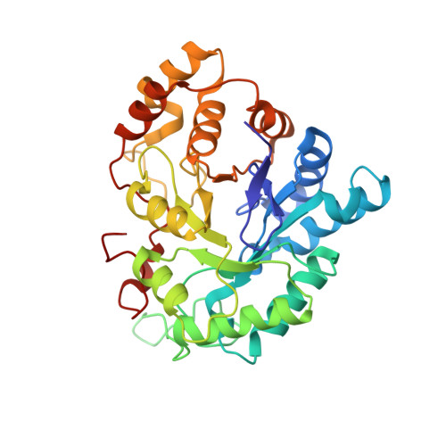
    3BCJ

  • PubMed Abstract: 

    The structure of human aldose reductase in complex with the 2 S4 R stereoisomer of the potent inhibitor Fidarestat ((2 S,4 S)-6-fluoro-2',5'-dioxospiro-[chroman-4,4'-imidazoline]-2-carboxamide) was determined at 15 K and a resolution of 0.78 A. The structure of the complex provides experimental evidence for the inhibition mechanism in which Fidarestat is initially bound neutral and then becomes negatively charged by donating the proton at the 1'-position nitrogen of the cyclic imide ring to the N2 atom of the catalytic His110.


  • Organizational Affiliation

    Department of Medicinal Chemistry, Monash University,Vic 3052, Australia.


Macromolecules
Find similar proteins by:  (by identity cutoff)  |  3D Structure
Entity ID: 1
MoleculeChains Sequence LengthOrganismDetailsImage
Aldose reductase316Homo sapiensMutation(s): 0 
EC: 1.1.1.21
UniProt & NIH Common Fund Data Resources
Find proteins for P15121 (Homo sapiens)
Explore P15121 
Go to UniProtKB:  P15121
PHAROS:  P15121
GTEx:  ENSG00000085662 
Entity Groups  
Sequence Clusters30% Identity50% Identity70% Identity90% Identity95% Identity100% Identity
UniProt GroupP15121
Sequence Annotations
Expand
  • Reference Sequence
Small Molecules
Ligands 3 Unique
IDChains Name / Formula / InChI Key2D Diagram3D Interactions
NAP
Query on NAP

Download Ideal Coordinates CCD File 
B [auth A]NADP NICOTINAMIDE-ADENINE-DINUCLEOTIDE PHOSPHATE
C21 H28 N7 O17 P3
XJLXINKUBYWONI-NNYOXOHSSA-N
FIS
Query on FIS

Download Ideal Coordinates CCD File 
D [auth A](2S,4R)-2-AMINOFORMYL-6-FLUORO-SPIRO[CHROMAN-4,4'-IMIDAZOLIDINE]-2',5'-DIONE
C12 H10 F N3 O4
WAAPEIZFCHNLKK-QPUJVOFHSA-N
CIT
Query on CIT

Download Ideal Coordinates CCD File 
C [auth A]CITRIC ACID
C6 H8 O7
KRKNYBCHXYNGOX-UHFFFAOYSA-N
Binding Affinity Annotations 
IDSourceBinding Affinity
FIS BindingDB:  3BCJ IC50: 1.10e+4 (nM) from 1 assay(s)
Experimental Data & Validation

Experimental Data

  • Method: X-RAY DIFFRACTION
  • Resolution: 0.78 Å
  • R-Value Free: 0.110 
  • R-Value Work: 0.100 
  • R-Value Observed: 0.105 
  • Space Group: P 1 21 1
Unit Cell:
Length ( Å )Angle ( ˚ )
a = 49.2α = 90
b = 66.642β = 91.67
c = 47.28γ = 90
Software Package:
Software NamePurpose
SHELXmodel building
SHELXL-97refinement
HKL-2000data collection
HKL-2000data reduction
SCALEPACKdata scaling
SHELXphasing

Structure Validation

View Full Validation Report


Ligand Structure Quality Assessment 


Entry History 

Deposition Data

Revision History  (Full details and data files)

  • Version 1.0: 2008-04-08
    Type: Initial release
  • Version 1.1: 2011-07-13
    Changes: Version format compliance
  • Version 1.2: 2023-11-01
    Changes: Data collection, Database references, Derived calculations, Refinement description