


Crystal structure of predicted acetamidase/formamidase (YP_546212.1) from Methylobacillus flagellatus KT at 1.58 A resolution
Joint Center for Structural Genomics (JCSG)To be published.
Experimental Data Snapshot
wwPDB Validation 3D Report Full Report

Entity ID: 1 | |||||
|---|---|---|---|---|---|
| Molecule | Chains | Sequence Length | Organism | Details | Image |
| Twin-arginine translocation pathway signal protein | 484 | Methylobacillus flagellatus KT | Mutation(s): 0 Gene Names: YP_546212.1, Mfla_2104 | 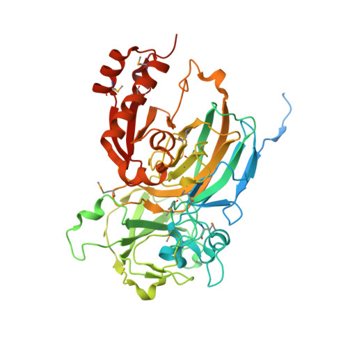 | |
UniProt | |||||
Find proteins for Q1GZG6 (Methylobacillus flagellatus (strain KT / ATCC 51484 / DSM 6875)) Explore Q1GZG6 Go to UniProtKB: Q1GZG6 | |||||
Entity Groups | |||||
| Sequence Clusters | 30% Identity50% Identity70% Identity90% Identity95% Identity100% Identity | ||||
| UniProt Group | Q1GZG6 | ||||
Sequence AnnotationsExpand | |||||
| |||||
| Ligands 3 Unique | |||||
|---|---|---|---|---|---|
| ID | Chains | Name / Formula / InChI Key | 2D Diagram | 3D Interactions | |
| EDO Query on EDO | AA [auth B] AB [auth D] BB [auth D] CB [auth D] DA [auth C] | 1,2-ETHANEDIOL C2 H6 O2 LYCAIKOWRPUZTN-UHFFFAOYSA-N |  | ||
| CL Query on CL | G [auth A], SA [auth D] | CHLORIDE ION Cl VEXZGXHMUGYJMC-UHFFFAOYSA-M |  | ||
| MG Query on MG | BA [auth C] CA [auth C] E [auth A] F [auth A] QA [auth D] | MAGNESIUM ION Mg JLVVSXFLKOJNIY-UHFFFAOYSA-N |  | ||
| Modified Residues 1 Unique | |||||
|---|---|---|---|---|---|
| ID | Chains | Type | Formula | 2D Diagram | Parent |
| MSE Query on MSE | A, B, C, D | L-PEPTIDE LINKING | C5 H11 N O2 Se |  | MET |
| Length ( Å ) | Angle ( ˚ ) |
|---|---|
| a = 62.165 | α = 107.82 |
| b = 82.723 | β = 105.81 |
| c = 83.576 | γ = 95.18 |
| Software Name | Purpose |
|---|---|
| REFMAC | refinement |
| PHENIX | refinement |
| SOLVE | phasing |
| MolProbity | model building |
| SCALA | data scaling |
| PDB_EXTRACT | data extraction |
| ADSC | data collection |
| MOSFLM | data reduction |
RCSB PDB (citation) is hosted by
RCSB PDB is a member of the

