3B96

Structural Basis for Substrate Fatty-Acyl Chain Specificity: Crystal Structure of Human Very-Long-Chain Acyl-CoA Dehydrogenase


Experimental Data Snapshot

  • Method: X-RAY DIFFRACTION
  • Resolution: 1.91 Å
  • R-Value Free: 0.216 
  • R-Value Work: 0.159 
  • R-Value Observed: 0.162 

wwPDB Validation   3D Report Full Report


Ligand Structure Quality Assessment 


This is version 1.3 of the entry. See complete history


Literature

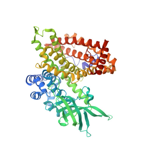
Structural basis for substrate fatty acyl chain specificity: crystal structure of human very-long-chain acyl-CoA dehydrogenase.

McAndrew, R.P.Wang, Y.Mohsen, A.W.He, M.Vockley, J.Kim, J.J.

(2008) J Biol Chem 283: 9435-9443

  • DOI: https://doi.org/10.1074/jbc.M709135200
  • Primary Citation of Related Structures:  
    3B96

  • PubMed Abstract: 

    Very-long-chain acyl-CoA dehydrogenase (VLCAD) is a member of the family of acyl-CoA dehydrogenases (ACADs). Unlike the other ACADs, which are soluble homotetramers, VLCAD is a homodimer associated with the mitochondrial membrane. VLCAD also possesses an additional 180 residues in the C terminus that are not present in the other ACADs. We have determined the crystal structure of VLCAD complexed with myristoyl-CoA, obtained by co-crystallization, to 1.91-A resolution. The overall fold of the N-terminal approximately 400 residues of VLCAD is similar to that of the soluble ACADs including medium-chain acyl-CoA dehydrogenase (MCAD). The novel C-terminal domain forms an alpha-helical bundle that is positioned perpendicular to the two N-terminal helical domains. The fatty acyl moiety of the bound substrate/product is deeply imbedded inside the protein; however, the adenosine pyrophosphate portion of the C14-CoA ligand is disordered because of partial hydrolysis of the thioester bond and high mobility of the CoA moiety. The location of Glu-422 with respect to the C2-C3 of the bound ligand and FAD confirms Glu-422 to be the catalytic base. In MCAD, Gln-95 and Glu-99 form the base of the substrate binding cavity. In VLCAD, these residues are glycines (Gly-175 and Gly-178), allowing the binding channel to extend for an additional 12A and permitting substrate acyl chain lengths as long as 24 carbons to bind. VLCAD deficiency is among the more common defects of mitochondrial beta-oxidation and, if left undiagnosed, can be fatal. This structure allows us to gain insight into how a variant VLCAD genotype results in a clinical phenotype.


  • Organizational Affiliation

    Department of Biochemistry, Medical College of Wisconsin, 8701 Watertown Plank Road, Milwaukee, WI 53226, USA.


Macromolecules
Find similar proteins by:  (by identity cutoff)  |  3D Structure
Entity ID: 1
MoleculeChains Sequence LengthOrganismDetailsImage
Very long-chain specific acyl-CoA dehydrogenase587Homo sapiensMutation(s): 0 
Gene Names: ACADVLVLCAD
EC: 1.3.99
UniProt & NIH Common Fund Data Resources
Find proteins for P49748 (Homo sapiens)
Explore P49748 
Go to UniProtKB:  P49748
PHAROS:  P49748
GTEx:  ENSG00000072778 
Entity Groups  
Sequence Clusters30% Identity50% Identity70% Identity90% Identity95% Identity100% Identity
UniProt GroupP49748
Sequence Annotations
Expand
  • Reference Sequence
Small Molecules
Ligands 2 Unique
IDChains Name / Formula / InChI Key2D Diagram3D Interactions
MYA
Query on MYA

Download Ideal Coordinates CCD File 
B [auth A]TETRADECANOYL-COA
C35 H62 N7 O17 P3 S
DUAFKXOFBZQTQE-QSGBVPJFSA-N
FAD
Query on FAD

Download Ideal Coordinates CCD File 
C [auth A]FLAVIN-ADENINE DINUCLEOTIDE
C27 H33 N9 O15 P2
VWWQXMAJTJZDQX-UYBVJOGSSA-N
Experimental Data & Validation

Experimental Data

  • Method: X-RAY DIFFRACTION
  • Resolution: 1.91 Å
  • R-Value Free: 0.216 
  • R-Value Work: 0.159 
  • R-Value Observed: 0.162 
  • Space Group: C 2 2 21
Unit Cell:
Length ( Å )Angle ( ˚ )
a = 74.616α = 90
b = 107.879β = 90
c = 149.776γ = 90
Software Package:
Software NamePurpose
DENZOdata reduction
SCALEPACKdata scaling
SOLVEphasing
DMphasing
REFMACrefinement
PDB_EXTRACTdata extraction

Structure Validation

View Full Validation Report


Ligand Structure Quality Assessment 


Entry History 

Deposition Data

Revision History  (Full details and data files)

  • Version 1.0: 2008-02-12
    Type: Initial release
  • Version 1.1: 2011-07-13
    Changes: Version format compliance
  • Version 1.2: 2017-10-25
    Changes: Refinement description
  • Version 1.3: 2024-02-21
    Changes: Data collection, Database references, Derived calculations