3B8Z

High Resolution Crystal Structure of the Catalytic Domain of ADAMTS-5 (Aggrecanase-2)


Experimental Data Snapshot

  • Method: X-RAY DIFFRACTION
  • Resolution: 1.40 Å
  • R-Value Free: 0.214 
  • R-Value Work: 0.185 
  • R-Value Observed: 0.187 

wwPDB Validation   3D Report Full Report


Ligand Structure Quality Assessment 


This is version 1.3 of the entry. See complete history


Literature

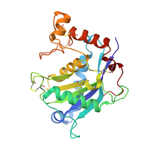
High resolution crystal structure of the catalytic domain of ADAMTS-5 (aggrecanase-2).

Shieh, H.S.Mathis, K.J.Williams, J.M.Hills, R.L.Wiese, J.F.Benson, T.E.Kiefer, J.R.Marino, M.H.Carroll, J.N.Leone, J.W.Malfait, A.M.Arner, E.C.Tortorella, M.D.Tomasselli, A.

(2008) J Biol Chem 283: 1501-1507

  • DOI: https://doi.org/10.1074/jbc.M705879200
  • Primary Citation of Related Structures:  
    3B8Z

  • PubMed Abstract: 

    Aggrecanase-2 (a disintegrin and metalloproteinase with thrombospondin motifs-5 (ADAMTS-5)), a member of the ADAMTS protein family, is critically involved in arthritic diseases because of its direct role in cleaving the cartilage component aggrecan. The catalytic domain of aggrecanase-2 has been refolded, purified, and crystallized, and its three-dimensional structure determined to 1.4A resolution in the presence of an inhibitor. A high resolution structure of an ADAMTS/aggrecanase protein provides an opportunity for the development of therapeutics to treat osteoarthritis.


  • Organizational Affiliation

    Pfizer Global Research and Development, St. Louis, Missouri 63017.


Macromolecules
Find similar proteins by:  (by identity cutoff)  |  3D Structure
Entity ID: 1
MoleculeChains Sequence LengthOrganismDetailsImage
protein ADAMTS-5
A, B
217Homo sapiensMutation(s): 1 
Gene Names: ADAMTS5ADAMTS11ADMP2
EC: 3.4.24
UniProt & NIH Common Fund Data Resources
Find proteins for Q9UNA0 (Homo sapiens)
Explore Q9UNA0 
Go to UniProtKB:  Q9UNA0
PHAROS:  Q9UNA0
GTEx:  ENSG00000154736 
Entity Groups  
Sequence Clusters30% Identity50% Identity70% Identity90% Identity95% Identity100% Identity
UniProt GroupQ9UNA0
Sequence Annotations
Expand
  • Reference Sequence
Small Molecules
Ligands 3 Unique
IDChains Name / Formula / InChI Key2D Diagram3D Interactions
294
Query on 294

Download Ideal Coordinates CCD File 
G [auth A],
L [auth B]
N-hydroxy-4-({4-[4-(trifluoromethyl)phenoxy]phenyl}sulfonyl)tetrahydro-2H-pyran-4-carboxamide
C19 H18 F3 N O6 S
FOSWRYKPHVPIDJ-UHFFFAOYSA-N
ZN
Query on ZN

Download Ideal Coordinates CCD File 
C [auth A],
H [auth B]
ZINC ION
Zn
PTFCDOFLOPIGGS-UHFFFAOYSA-N
CA
Query on CA

Download Ideal Coordinates CCD File 
D [auth A]
E [auth A]
F [auth A]
I [auth B]
J [auth B]
D [auth A],
E [auth A],
F [auth A],
I [auth B],
J [auth B],
K [auth B]
CALCIUM ION
Ca
BHPQYMZQTOCNFJ-UHFFFAOYSA-N
Binding Affinity Annotations 
IDSourceBinding Affinity
294 PDBBind:  3B8Z IC50: 290 (nM) from 1 assay(s)
Binding MOAD:  3B8Z IC50: 290 (nM) from 1 assay(s)
Experimental Data & Validation

Experimental Data

  • Method: X-RAY DIFFRACTION
  • Resolution: 1.40 Å
  • R-Value Free: 0.214 
  • R-Value Work: 0.185 
  • R-Value Observed: 0.187 
  • Space Group: P 1 21 1
Unit Cell:
Length ( Å )Angle ( ˚ )
a = 53.057α = 90
b = 44.487β = 90.07
c = 76.38γ = 90
Software Package:
Software NamePurpose
DENZOdata reduction
SCALEPACKdata scaling
AMoREphasing
REFMACrefinement
PDB_EXTRACTdata extraction
ADSCdata collection
HKL-2000data reduction
HKL-2000data scaling

Structure Validation

View Full Validation Report


Ligand Structure Quality Assessment 


Entry History 

Deposition Data

Revision History  (Full details and data files)

  • Version 1.0: 2007-12-11
    Type: Initial release
  • Version 1.1: 2011-07-13
    Changes: Version format compliance
  • Version 1.2: 2017-10-25
    Changes: Refinement description
  • Version 1.3: 2021-10-20
    Changes: Database references, Derived calculations