


The crystal structure of the cytidine deaminase from Bacillus anthracis.
Zhang, R., Joachimiak, G., Wu, R., Patterson, S., Gornicki, P., Joachimiak, A.To be published.
Experimental Data Snapshot
wwPDB Validation 3D Report Full Report

Entity ID: 1 | |||||
|---|---|---|---|---|---|
| Molecule | Chains | Sequence Length | Organism | Details | Image |
| Putative Blasticidin S deaminase | 142 | Bacillus anthracis | Mutation(s): 0 Gene Names: BAS3426, BA_3696, GBAA3696 EC: 3.5.4.4 | 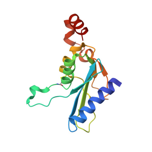 | |
UniProt | |||||
Find proteins for A0A6H3AIE5 (Bacillus anthracis) Explore A0A6H3AIE5 Go to UniProtKB: A0A6H3AIE5 | |||||
Entity Groups | |||||
| Sequence Clusters | 30% Identity50% Identity70% Identity90% Identity95% Identity100% Identity | ||||
| UniProt Group | A0A6H3AIE5 | ||||
Sequence AnnotationsExpand | |||||
| |||||
| Length ( Å ) | Angle ( ˚ ) |
|---|---|
| a = 57.078 | α = 90 |
| b = 98.479 | β = 111.09 |
| c = 61.36 | γ = 90 |
| Software Name | Purpose |
|---|---|
| REFMAC | refinement |
| SBC-Collect | data collection |
| HKL-3000 | data reduction |
| HKL-3000 | data scaling |
| HKL-3000 | phasing |
RCSB PDB (citation) is hosted by
RCSB PDB is a member of the

